A curious nose. A very waggy welcome.
If your vet has recommended an echocardiogram for your dog, you might be picturing something intense. Totally fair. A cardiology visit can feel like a big step. The good news is that an echo is usually calm, noninvasive, and packed with useful information. It’s one of the best ways to look at the heart in real time and figure out what is going on with murmurs, coughing, fainting episodes, or suspected heart disease.
What a dog echocardiogram is
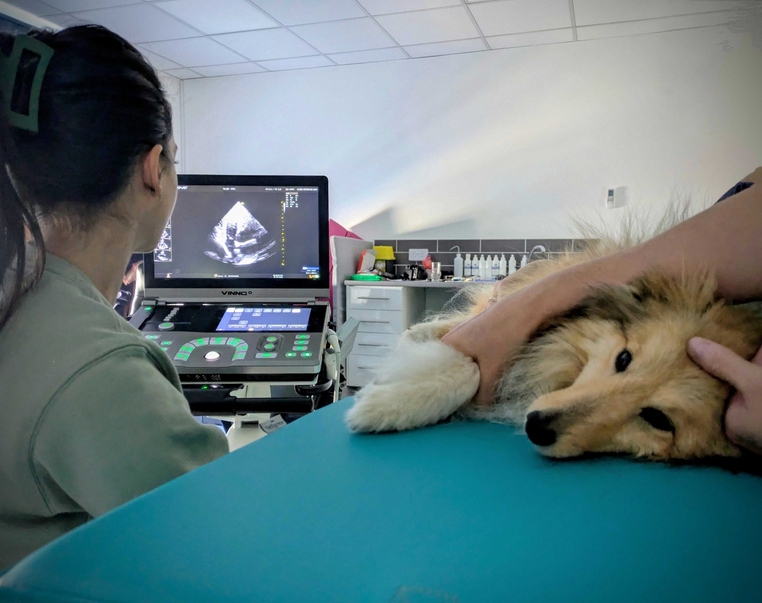
An echocardiogram is a cardiac ultrasound. It uses sound waves to create moving images of your dog’s heart, including the chambers, valves, and major vessels leaving and entering the heart. Unlike X-rays, an echo does not use radiation. And unlike an ECG (electrocardiogram), which focuses on electrical rhythm, an echo focuses on structure and function.
Many general practices can do basic ultrasounds, but a full heart echo is often performed by a veterinary cardiologist or a hospital with a cardiology service. The person doing the scan matters because measurements and interpretation guide long-term decisions like when to start medication and which drugs make sense.
What an echocardiogram measures
Think of an echo like a very detailed map of how the heart is built and how it moves blood. Common things an echocardiogram can evaluate include:
- Heart chamber size (for example, whether the left atrium is enlarged)
- Wall thickness (important when screening for certain diseases or high blood pressure effects)
- Pumping strength and overall function
- Valve structure (whether a valve looks thickened or prolapsing)
- Valve leaks or abnormal blood flow using Doppler ultrasound (often described as regurgitation)
- Pressure estimates that can suggest pulmonary hypertension in some cases
- Fluid around the heart (pericardial effusion)
- Masses or unusual anatomy (less common, but important to catch)
This is why echoes are so closely tied to conditions like myxomatous mitral valve disease (MMVD). A murmur can tell you something is happening, but an echo can help show how much the valve is leaking, whether the heart is remodeling, and whether the situation is stable or heading toward congestive heart failure.
What to expect at the appointment
Check-in and a quick history
You will usually be asked about symptoms like coughing, exercise intolerance, fast breathing during sleep, fainting, appetite changes, or belly swelling. Bring a list of current medications and supplements, plus any recent lab work or imaging your regular vet has done.
Prep: a small clipped patch is common
Most dogs need a small area of fur clipped on the chest so the ultrasound probe can make clean contact with the skin. Ultrasound gel is applied. This is messy but harmless, and it wipes off easily.
Positioning: often lying on a special table
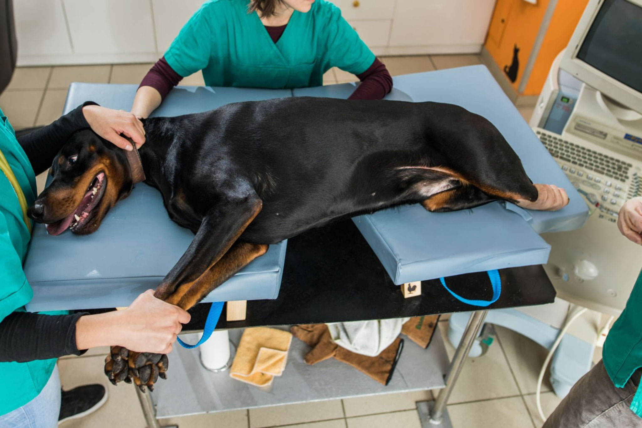
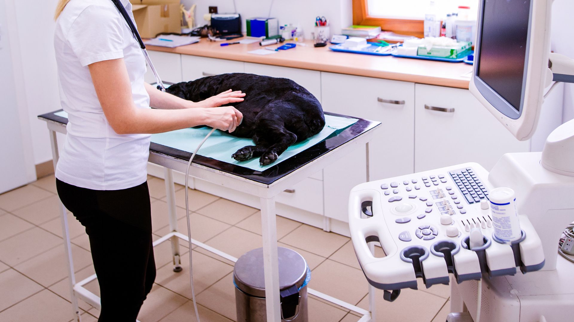
Many cardiology clinics use a padded echo table with a cutout so the probe can be placed against the chest from underneath. Dogs are typically gently positioned on their side. A technician may help keep your pup steady and comfortable.
During the scan: quiet, focused, and patient
The scan itself is commonly 30 to 60 minutes, but it can be shorter for screening studies or longer for complex cases, wiggly dogs, or congenital disease evaluations. The cardiologist will capture still images and video loops and take specific measurements.
Afterward: discussion and a plan
Many clinics review findings with you the same day. In some situations, you may receive a written report later or your primary veterinarian will get the full report with measurements and recommendations.
Does a dog need sedation for an echocardiogram?
Often, no. Most dogs can have an echocardiogram while awake. The goal is to keep them calm and still so the images are accurate. Many clinics avoid sedation unless it is truly needed, because some sedatives can affect heart rate and blood pressure, which can influence interpretation.
That said, mild sedation is sometimes used on purpose to reduce stress and wiggling, especially for very anxious dogs. If sedation is used, your veterinary team will choose drugs and doses carefully based on your dog’s health status and the information they need from the study.
If you are worried about stress, ask ahead what comfort options they offer. Sometimes the simplest tools help most, like a quiet room, a slow approach, a non-slip mat, and a calm handler.
How to prepare your dog
- Ask about fasting. Many echoes do not require fasting, but policies vary, especially if sedation is possible.
- Bring favorite treats if allowed. Tiny, soft treats can help nervous dogs settle.
- Keep your dog’s harness and leash simple so the chest area is easy to access.
- Bring records from your primary vet, including murmur grade notes, X-rays, bloodwork, and medication history.
- Plan for time. Specialty appointments can run longer than you expect, especially in busy hospitals.
If your dog is already on heart medication, do not stop it unless your veterinarian or cardiologist specifically tells you to.
How much it costs
Echocardiogram pricing varies a lot based on location, whether a board-certified cardiologist performs the study, and what is included in the visit. The ranges below reflect a typical U.S. specialty setting. Costs may be lower at community screening clinics and can be higher in high-cost areas, urgent visits, or complex cases.
- Typical U.S. specialty range: about $500 to $1,200 for an echocardiogram performed through a specialty hospital or cardiology practice.
- Lower end may be seen for limited studies, rechecks, or screening clinics.
- Higher end is more common in high-cost metro areas, emergency settings, or cases that require additional monitoring or add-on testing.
Possible add-on costs that can change the total include:
- Cardiology consultation or specialist exam fee
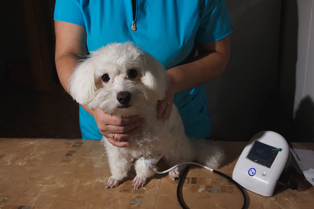
- Blood pressure measurement
- Chest X-rays (radiographs)
- ECG, Holter monitor, or rhythm monitoring
- NT-proBNP blood test or other lab work (case-dependent)
- Sedation (if needed)
For a more accurate estimate, ask the clinic for an itemized quote. If cost is a big concern, tell them. Some practices can discuss what is most essential now versus what can wait.
Other tests you might see
It is very common for a cardiology visit to include more than just the echo. These add-ons are not “busywork.” They answer different questions.
- Chest X-rays: show the heart’s overall silhouette and the lungs. They can help identify cardiac enlargement patterns and signs of pulmonary edema (fluid in the lungs), but they cannot show valve motion or blood flow directly.
- ECG: checks rhythm and conduction. Some rhythm problems come and go, so a quick in-clinic ECG may be paired with longer monitoring.
- Holter or event monitor: tracks intermittent arrhythmias over hours or days, especially for fainting episodes.
- Blood pressure: helps interpret heart changes and can matter for treatment decisions.
What an echo cannot tell you
An echocardiogram is powerful, but it is not a one-test answer to everything. If your dog is coughing, for example, an echo can help rule in or rule out heart disease as the main driver, but coughing can also be caused by airway or lung disease that needs different diagnostics. And while an echo can hint at rhythm issues, many arrhythmias require an ECG or longer monitoring to fully understand.
How results guide treatment
An echocardiogram is not just about naming a condition. It helps your veterinary team decide what to do next and when to do it. Depending on what the echo shows, that plan may include:
- Monitoring only (for example, a mild murmur with no meaningful heart enlargement)
- Starting or adjusting medication to reduce workload on the heart, manage fluid, or address rhythm issues
- Setting recheck timing (some dogs need echoes every 6 to 12 months, others sooner)
- Exercise guidance tailored to your dog’s current heart function
- Anesthesia risk planning for dental cleanings or other procedures
For MMVD specifically, echo measurements help determine staging and risk. Some dogs with heart enlargement from MMVD (often called Stage B2) may benefit from medication aimed at slowing progression and reducing the likelihood of developing congestive heart failure. Dogs without enlargement typically do not need that same approach yet, even if a murmur is present.
If you take only one thing from this page, let it be this: a murmur is a clue, but an echocardiogram is the roadmap.
When an echocardiogram is recommended
Your vet may suggest an echo if your dog has:
- A newly discovered heart murmur
- A murmur that has gotten louder over time
- Coughing, especially at night or after activity
- Fast or difficult breathing
- Exercise intolerance or tiring quickly on walks
- Fainting or sudden weakness episodes
- Abnormal heart rhythm
- Breed risk factors (especially small breeds prone to valve disease)
An echo may also be recommended before anesthesia in some dogs with known murmurs, particularly if the murmur is moderate to loud, the dog is older, or there are any symptoms.
Urgent signs to not ignore
If your dog is struggling to breathe (open-mouth breathing at rest, severe effort, blue or gray gums, collapse, or sudden extreme weakness), treat it as an emergency. Do not wait for a scheduled echocardiogram appointment. Seek urgent veterinary care right away.
At-home monitoring
If your dog has a murmur or diagnosed heart disease, ask your vet if they want you to track resting respiratory rate. When your dog is asleep or deeply relaxed, count breaths for 30 seconds and multiply by two. A consistent upward trend matters more than a single reading, so write it down and bring the log to rechecks.
Questions to ask
- Is this a full echocardiogram with Doppler, or a focused scan?
- Will a cardiologist interpret the study?
- Do you expect my dog will need sedation?
- What stage is the disease, and what does that mean day to day?
- What changes should I watch for at home (resting breathing rate, cough, energy)?
- When should we recheck, and what would trigger an earlier visit?
- How will this affect anesthesia safety for dental work or surgery?
FAQ
Is an echocardiogram painful?
It should not be painful. The probe is gently pressed against the chest with gel. The most annoying part for many dogs is the clipping and the need to lie still.
How long does it take?
Many appointments include a physical exam, the scan, and a discussion of results. The echo portion is often 30 to 60 minutes, but it varies based on the type of study and how cooperative your dog feels that day.
Will I get results the same day?
Often yes, at least a summary. Some clinics send a detailed written report to your primary vet and may follow up with you once the full measurements are finalized.
What is the difference between an X-ray and an echocardiogram?
X-rays show the heart’s overall outline and the lungs and can help identify patterns of heart enlargement and lung changes like pulmonary edema. An echocardiogram shows the inside of the heart, including valves, blood flow direction, and pumping function. They answer different questions and are often used together.
How often will my dog need an echocardiogram?
It depends on the diagnosis and stage. Some dogs only need periodic monitoring, while others need rechecks more frequently to fine-tune medication or watch for progression.