
When people hear “fluid,” they often think of fluid in the lungs or a swollen belly. But there is another kind of fluid that can be even more immediately dangerous: fluid around the heart. In dogs, that is called pericardial effusion, and it is one of those conditions where time truly matters.
As a veterinary assistant here in Frisco, Texas, I have seen how fast a dog can go from “seems a little off” to a true emergency when that fluid starts squeezing the heart. The good news is that with quick care, many dogs can stabilize. The key is recognizing the signs and getting to a veterinarian right away.
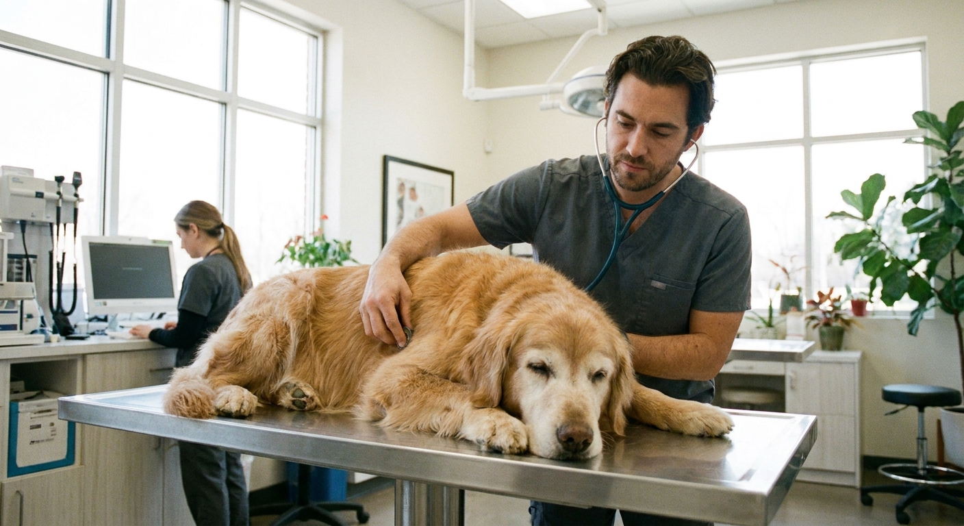
What is pericardial effusion?
The heart sits inside a thin, protective sac called the pericardium. Normally, there is only a tiny amount of lubricating fluid in that space.
Pericardial effusion means there is an abnormal buildup of fluid inside that sac. The problem is not just the fluid itself. It is what the fluid does. As it accumulates, it can compress the heart so the chambers cannot fill normally between beats.
When the heart cannot fill, it cannot pump enough blood to the body. This can lead to poor perfusion, shock, and collapse.
Why this is an emergency: cardiac tamponade
When pericardial fluid builds up enough to significantly restrict the heart’s ability to fill, veterinarians call it cardiac tamponade.
In plain language, tamponade is when the heart is being “squeezed” from the outside. A dog can look weak, breathe hard, or suddenly collapse because the body is not getting enough blood flow.
If you suspect pericardial effusion, it is safest to treat it as an emergency, even if your dog seems to perk up again.
Signs of pericardial effusion
Some dogs show subtle signs at first, especially if the fluid accumulates gradually. Others crash quickly.
Signs you may notice at home
- Collapse or fainting episodes
- Weakness, reluctance to move, or sudden exercise intolerance
- Rapid breathing, panting, or shallow breaths that feel “off” for your dog
- Labored breathing or working harder to breathe
- Pale gums (or gums that look grayish)
- Restlessness and inability to get comfortable
- Swollen belly that can develop over time (fluid backup can cause abdominal enlargement, especially with slower or recurring effusion)
- Reduced appetite or seeming “not themselves”
Signs your vet may find on exam
- Muffled heart sounds (the fluid dampens what we hear with a stethoscope)
- Weak pulses and sometimes pulsus paradoxus (pulses can feel weaker with breathing in)
- Fast heart rate
- Jugular vein distension (neck veins may look more prominent)
- Low blood pressure and poor circulation
One important point: some dogs with pericardial effusion do not have an obvious heart murmur. That can surprise families who assume “heart trouble” always equals a murmur.
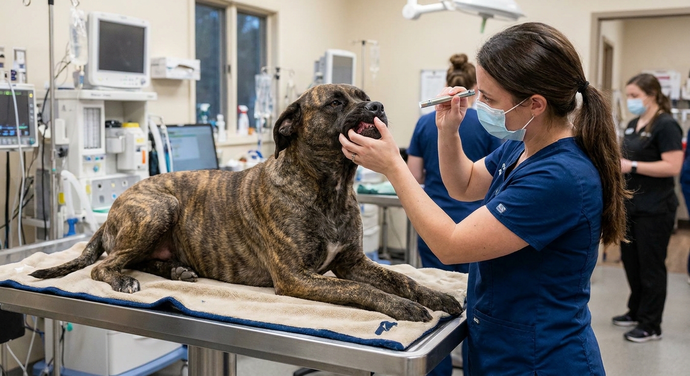
What causes fluid around the heart?
Pericardial effusion is a finding, not a single disease. The fluid can be blood, inflammatory fluid, or less commonly other types of fluid. Your vet’s job is to determine why it is happening, because that affects treatment and prognosis.
Idiopathic pericardial effusion
Idiopathic means we do not find a clear underlying cause, even after a reasonable workup. This can happen in dogs, and some respond well to draining the fluid. In some cases, the effusion comes back and further procedures may be discussed.
Bleeding from a heart mass
Another common scenario is bleeding into the pericardial sac from a mass in or near the heart. The classic location is the right atrium (for example with hemangiosarcoma) or the heart base (for example chemodectoma, ectopic thyroid carcinoma, or other tumors). Mesothelioma can also involve the pericardium. The fluid is often bloody.
Some breeds are overrepresented with certain causes (you may hear vets mention Golden Retrievers, German Shepherds, and other large-breed dogs), but any dog can be affected.
This is one reason pericardial effusion can be so sudden and severe: a bleeding event can rapidly increase pressure around the heart.
Other possible causes
- Inflammation (pericarditis), and less commonly infection
- Clotting disorders or anticoagulant rodenticide exposure (can cause bleeding)
- Trauma (for example, being hit by a car)
- Other tumors and underlying diseases
Because the causes vary so much, it is very normal for your veterinarian to recommend imaging and lab work fairly quickly once your dog is stable.
Effusion vs a heart murmur visit
A lot of families have already been through an appointment for a “routine” murmur. That visit often focuses on grading how loud the murmur is, discussing whether an echocardiogram is needed, and checking for signs of chronic progression.
Pericardial effusion is different. It is not primarily about murmur intensity. It is about whether the heart is being compressed and whether the dog is in shock.
How the approach is different
- Priority is stabilization: oxygen, IV access, careful fluid support, and rapid assessment.
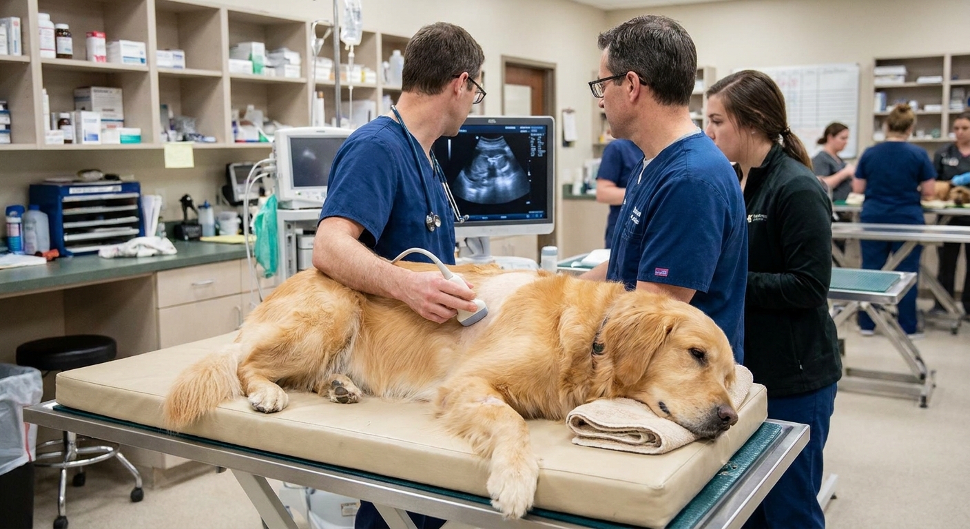
- Bedside ultrasound is a game-changer: many ER teams use a quick focused ultrasound to confirm fluid around the heart in minutes.
- An emergency procedure may be needed: draining the fluid can be life-saving.
- Cause investigation comes after: once circulation improves, your vet can better discuss next diagnostic steps.
How vets diagnose it
Diagnosis usually involves a combination of physical exam findings and imaging.
Tests commonly used
- Focused ultrasound or echocardiogram to see fluid around the heart and evaluate how well the heart is filling
- Chest X-rays to assess heart silhouette and lungs (X-rays can suggest effusion, but ultrasound is more definitive)
- Electrocardiogram (ECG) to check rhythm and look for patterns that can occur with effusion
- Blood pressure and perfusion checks
- Bloodwork (CBC/chemistry) to evaluate anemia, organ function, and overall stability
- Clotting tests if bleeding risk or toxin exposure is suspected
Sometimes fluid is also collected and analyzed. Fluid analysis can help narrow down inflammation versus bleeding, but it does not always reveal a single clear answer.
What happens in the ER
If your dog comes in unstable, the first goal is to support circulation and confirm what we are dealing with. In many ERs, this moves quickly.
- Triage and oxygen if breathing is hard or gums are pale
- IV catheter placement and careful support based on blood pressure and perfusion
- Focused ultrasound to look for pericardial fluid and signs of tamponade
- Monitoring for blood pressure changes and heart rhythm issues
- Pericardiocentesis if tamponade is present or suspected
It can look dramatic, but the fast pace is intentional. Once the pressure around the heart is relieved, many dogs visibly perk up.
Treatment: pericardiocentesis
The most immediate, life-saving treatment for cardiac tamponade is often pericardiocentesis, which is a procedure where a veterinarian uses a sterile needle and catheter to drain fluid from the pericardial sac.
This is typically done with ultrasound guidance and careful monitoring. Many dogs improve quickly once the pressure is relieved because the heart can fill normally again.
Risks and considerations
Every procedure has risks. With pericardiocentesis, your veterinarian may mention concerns like temporary arrhythmias, bleeding, accidental injury to nearby structures, or re-accumulation of fluid. The reason we still do it when indicated is simple: untreated tamponade can be fatal.
Other treatments your dog may need
- Oxygen therapy
- IV catheter placement and medications as needed
- Pain control or mild sedation depending on stability
- Treatment for underlying causes if identified (for example, addressing a clotting problem)
If it comes back
If the effusion recurs, your veterinarian may discuss referral to a cardiologist or specialty hospital. Depending on the suspected cause and your dog’s overall condition, options can include repeat drainage, advanced imaging, and in selected cases surgery such as a pericardial window or pericardiectomy.
What to do at home
If your dog collapses, seems suddenly weak, has very pale gums, or has a swollen belly with labored breathing, it is safest to assume this could be an emergency.
Steps while you head to care
- Go to an emergency veterinarian immediately. Call on the way if possible so they can prepare.
- Keep your dog calm and still. Excitement and exertion can worsen collapse risk.
- Do not force food, water, or medications unless a veterinarian instructs you to, including human pain medications.
- Carry small dogs. For larger dogs, use a blanket as a stretcher if they cannot walk safely.
If your dog is having trouble breathing or collapses, do not “wait and see.” Pericardial effusion can look like it improves briefly, then deteriorates again.
Prognosis
The honest answer is: it depends on the cause, how much fluid is present, and how quickly treatment happens.
- Idiopathic effusion often has a better outlook, especially if no mass is found and the effusion does not recur.
- Recurrent effusion may mean additional procedures or specialist care are needed.
- Bleeding from a mass can carry a more guarded prognosis, and your veterinarian will guide you through realistic next steps once your dog is stable.
What I want you to take away is this: even when the long-term cause is serious, urgent care can still buy you time and, in many cases, meaningful good days with your dog.
FAQ
Can pericardial effusion happen without a heart murmur?
Yes. Many dogs with pericardial effusion do not have a noticeable murmur. In fact, muffled heart sounds are often more typical than a loud murmur because the fluid dampens what we hear.
Does a swollen belly always mean fluid around the heart?
No. A swollen belly can come from many causes. With pericardial effusion, abdominal distension can occur because blood flow returning to the heart backs up, leading to fluid accumulation in the abdomen. But liver disease, internal bleeding, gastrointestinal issues, and other conditions can cause similar belly changes.
Is pericardial effusion the same as congestive heart failure?
No. Congestive heart failure means fluid buildup due to the heart not pumping efficiently. It often shows up as fluid in the lungs (left-sided failure) or fluid in the chest or abdomen (right-sided failure). Pericardial effusion is fluid in the sac around the heart that prevents normal filling. They are different problems and are managed differently.
How fast can this happen?
It can develop gradually over days to weeks, or it can worsen suddenly, especially if bleeding occurs. That unpredictability is why veterinarians treat suspected pericardial effusion as urgent.
Takeaway
Pericardial effusion in dogs is fluid in the sac around the heart that can prevent the heart from filling and pumping blood. The classic clues include muffled heart sounds, weak pulses, belly swelling, weakness, and collapse.
If you see these signs, please do not wait for a “regular appointment.” Head to an emergency veterinarian. Fast diagnosis and, when needed, drainage of the fluid can be life-saving.