When I’m helping families in the exam room, one of the hardest phone calls is the one that starts with: “The tumor came back as a mast cell tumor… and the margins weren’t clean.” Even when the lump is already gone, the word incomplete can make it feel like you are right back at square one.
Take a breath. An incompletely removed mast cell tumor (MCT) does not automatically mean your dog is out of options or that the cancer has already spread. It means we have to make a thoughtful plan based on what the pathology report actually says, what your dog’s tumor type looks like under the microscope, and what we can realistically do at that location on the body.
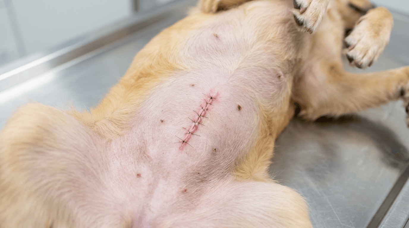
What “dirty margins” means
After your vet removes an MCT, the tissue is typically sent to a veterinary pathologist. One key job of the pathologist is to evaluate the surgical margins, meaning the edges of the removed tissue.
Clean margins
Clean margins means no tumor cells were seen right at the cut edges of the submitted tissue. In everyday terms: the pathologist did not see evidence that tumor was left behind at the edges of what was removed.
It is still not a guarantee that a tumor will never recur. Pathology is powerful, but it is not a crystal ball.
Dirty margins
Dirty margins (also described as incomplete excision or tumor extends to the margin) means tumor cells were reported at the edge of the evaluated tissue. The practical takeaway is that there is a higher chance that some microscopic tumor cells remain at the surgical site.
One nuance that matters: margin evaluation depends on how the specimen is handled and reported. Some labs use different trimming methods (for example, “radial” sections versus “en face” or “shaved” margins), and that can affect how margins are interpreted. Your veterinarian can tell you which lab was used and how they tend to report margins.
Close margins
Close or narrow margins usually means tumor cells were not seen at the exact edge, but they were seen very near it. The recurrence risk depends on tumor features and location.
A key detail owners often miss
Margin status is based on what was submitted and evaluated, not the entire surgical field in your dog’s body. Pathology is incredibly valuable, but it is still a sampling of tissue processed into thin slices. That is one reason vets talk in probabilities, not guarantees.
Report wording, translated
Here are phrases I see commonly, and what they typically mean in everyday language. If you have your report in hand, you may spot one of these exact lines.
- “Neoplastic cells extend to the margin”: tumor cells were reported at the cut edge, so residual microscopic disease is possible.
- “Margins are incomplete”: another way to say dirty margins.
- “Tumor present at deep margin”: the closest edge is the underside of the sample, meaning cells may remain deeper in the tissue bed. This is often harder to address than a superficial skin edge.
- “Tumor present at lateral margin”: the closest edge is one of the side margins, meaning cells may remain to one side of the scar.
- “Clean but narrow (X mm)”: no tumor at the very edge, but the buffer is small. Whether that is acceptable depends on tumor biology and location.
- “Cautery artifact”: heat from surgery made some areas hard to evaluate. This can limit margin interpretation.
- “Inflammation consistent with biopsy site”: normal healing changes where the lump was sampled or manipulated.
Most reports include other clues beyond grade, such as mitotic activity, whether the tumor is described as infiltrative, or comments about ulceration. These details help your veterinarian and any oncology specialist estimate the odds of local regrowth and spread.
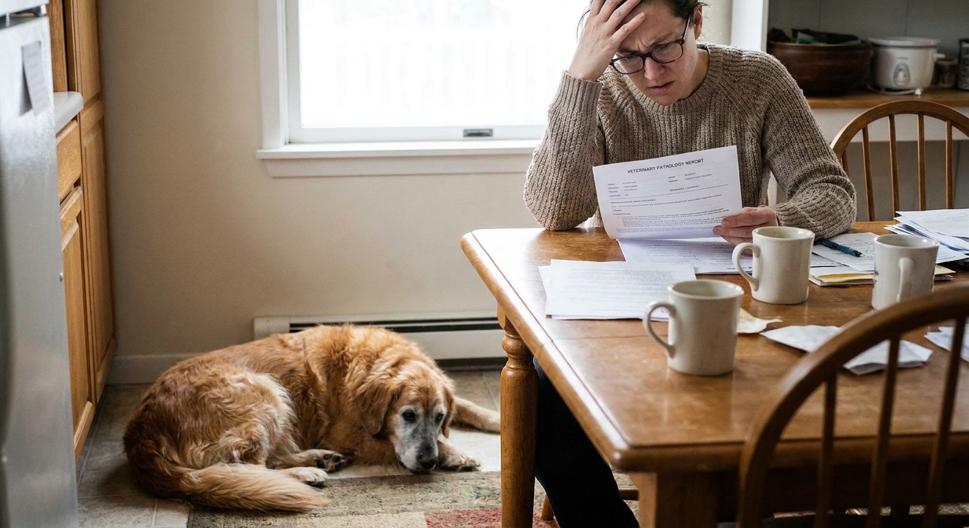
Grade terms can be confusing
Owners often hear “low grade” and assume all labs mean the same thing. Two grading systems are commonly referenced:
- Patnaik grade (I, II, III): an older 3-tier system. Many tumors were labeled Grade II, which is part of why it can feel less clear to owners.
- Kiupel grade (low or high): a newer 2-tier system that is often easier to apply clinically. When you hear “low-grade MCT,” it is frequently Kiupel low grade.
When I review a report with a family, I ask which system was used, and then I look at the other behavior markers on the page, not just the grade label.
Why margins are tricky for MCTs
Mast cell tumors can be infiltrative, sending microscopic extensions into surrounding tissues. What you see and feel as a lump is not always the full footprint. They also can look like they shrink and swell, because mast cells release histamine and other inflammatory chemicals.
That is why a surgeon can remove what looks like a generous amount of tissue, yet the pathology still comes back with incomplete edges.
First question: is anything visible?
Sometimes the “incomplete” finding is only microscopic. The incision can look perfect and flat, and there is no obvious lump. Other times, the surgeon already suspected the edges were tight because of the tumor’s location, or because the mass was more extensive than expected.
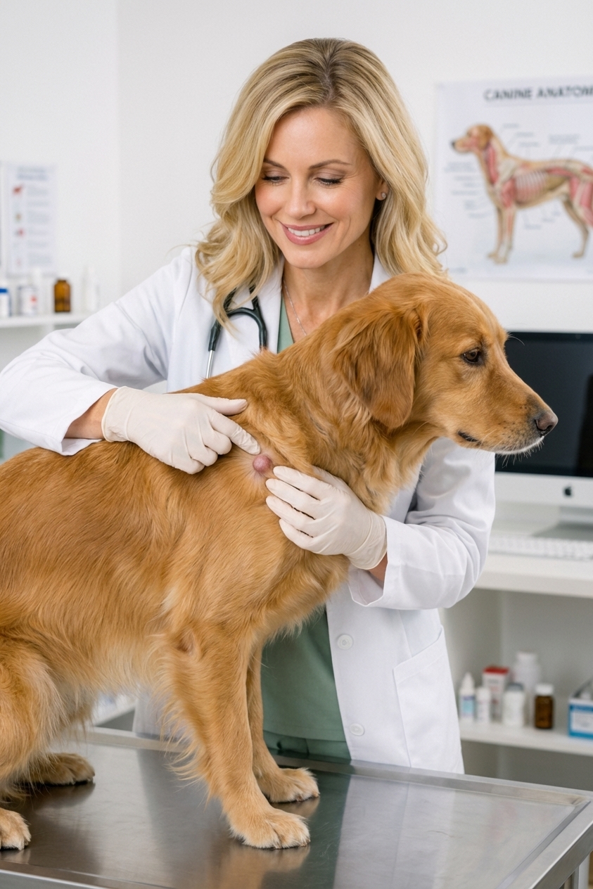
Your vet will usually do a focused recheck to assess:
- How the incision is healing
- Whether thickening feels like normal scar tissue or suspicious regrowth
- Whether the tumor location has enough extra skin and tissue for another attempt
This visit is also the moment to talk about next steps and referral options, because timing can matter if you plan a second surgery or radiation.
Normal scar or regrowth?
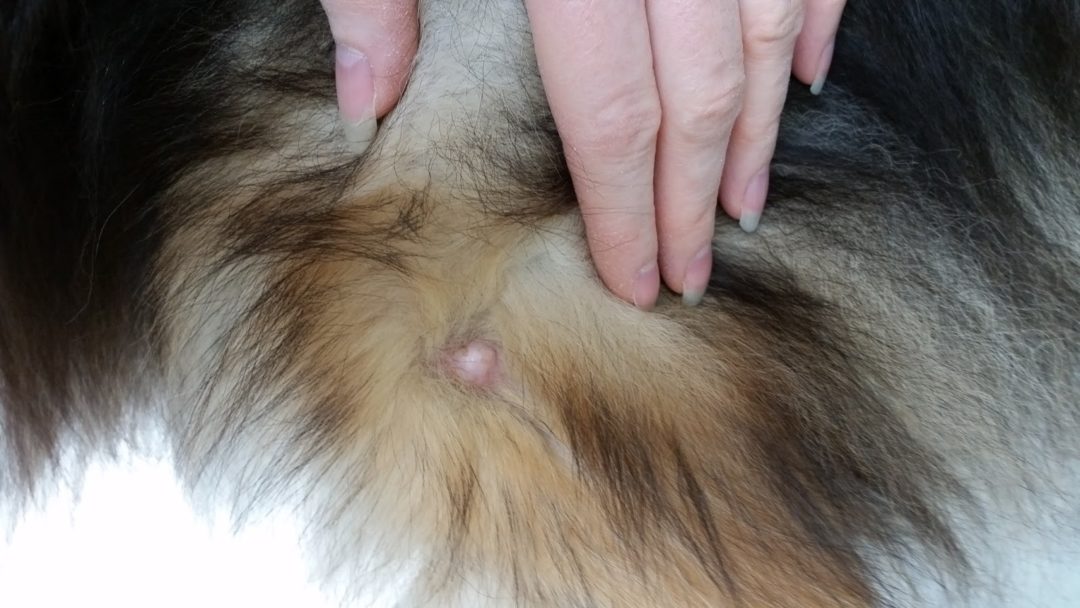
This is one of the most stressful parts for owners, and it is where clear expectations help. It is common for a healing incision to feel firmer or a little rope-like as scar tissue forms. Mild thickening can increase over the first few weeks and then slowly soften over time.
Things I consider more concerning include a new, discrete bump that seems to enlarge over days to weeks, a persistent bruised look, new redness and swelling that does not fit the healing timeline, or itchiness focused on the scar area. If you feel unsure, send your vet a photo and schedule a recheck. A quick look and feel can save you weeks of worry.
When re-excision is next
Re-excision (a second surgery to widen margins, sometimes called scar revision) means going back to remove more tissue around and under the previous surgery site, aiming for wider and deeper margins.
Re-excision is often favored when:
- Your dog is a good anesthesia candidate
- The tumor is in a location where wider margins are realistically possible (for example, trunk versus tight skin on lower legs)
- The pathology suggests the tumor is likely to behave locally if any cells remain
- You want the best shot at local control without radiation
Why timing matters
Many surgeons prefer re-excision before dense scar tissue forms, but your dog also needs enough time to heal. The exact timing is individualized. If you are considering a second surgery, ask: “If we plan re-excision, when would you ideally do it?”
What to ask the surgeon
- Which margin was incomplete: lateral (side) or deep (underneath)?
- How much additional tissue do you think you can safely remove at this site?
- Would you recommend referral to a board-certified surgeon for the second attempt?
When radiation comes next
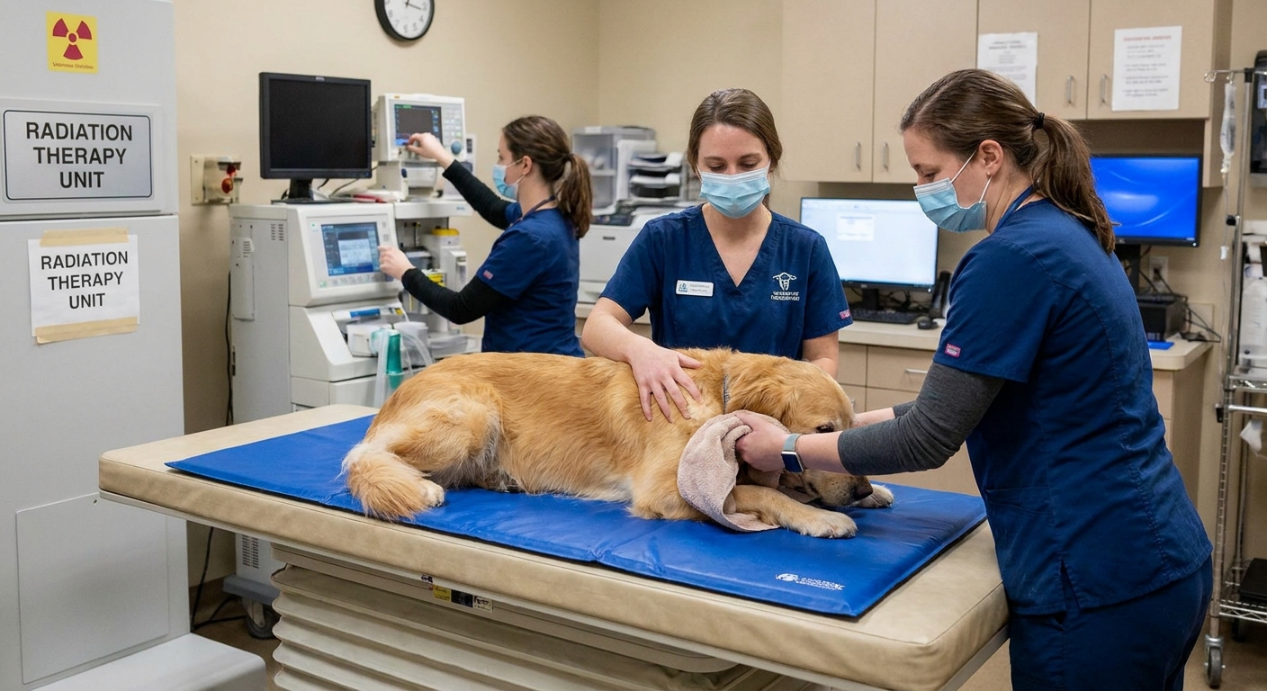
Radiation therapy is commonly used when an MCT was removed but microscopic disease is suspected to remain, and getting clean margins with another surgery would be difficult or overly invasive.
Radiation is often considered when:
- The tumor is in a challenging spot (lower leg, paw, face, around joints, or other tight areas)
- The report indicates incomplete margins and re-excision would not likely achieve a good buffer
- The deep margin is involved and going deeper would mean sacrificing important structures
- Your dog has already had one or more surgeries in that area
What a radiation consult covers
A radiation oncologist will review the pathology report, examine the site, and discuss whether they are treating microscopic residual cells (common after incomplete excision) or a measurable tumor. They will also explain expected side effects at that location and how many treatments are typical.
If you are on the fence, I still encourage owners to schedule the consult. A consult does not obligate you to start radiation. It simply gives you a clear, personalized plan.
Do we need staging?
“Staging” means checking for evidence of spread and clarifying overall risk. Not every dog needs every test, but incomplete margins are a good reason to pause and ask what else should be evaluated.
Common staging steps your vet or oncologist may discuss include:
- Regional lymph node sampling (often a needle aspirate), even if the node feels normal. Mast cell spread can be subtle early on.
- Basic bloodwork to make sure your dog is a safe anesthesia and treatment candidate.
- Abdominal ultrasound and, in higher-risk cases, sampling the spleen or liver. This is discussed more often for higher-grade tumors or worrisome pathology features.
Some pathology reports also mention, or your oncologist may recommend, additional risk markers (such as c-KIT staining pattern, KIT mutation testing, or proliferation markers like Ki-67). These tests are not required for every case, but they can be helpful when the next step feels unclear.
Palladia and other meds
Palladia (toceranib) is a targeted anti-cancer medication that is sometimes used for mast cell tumors, especially when disease is not easily controlled with local treatments alone.
The plain-language difference is this:
- Re-excision or radiation is primarily about local control, meaning preventing the tumor from regrowing at the scar site.
- Systemic therapy is about cancer behavior in the body more broadly. Palladia is one systemic option, and depending on grade and stage, oncologists may also discuss other chemotherapy protocols (for example vinblastine with prednisone, or lomustine/CCNU).
Why Palladia is not automatically first for low-risk cases
If the tumor appears low-grade and the main issue is microscopic cells potentially left in one spot, many dogs benefit most from addressing that spot directly with re-excision or radiation, rather than jumping straight to long-term medication.
Palladia can be a very useful tool, but it requires careful monitoring and can cause side effects. Your vet or oncologist weighs the risk of local recurrence and the risk of spread against the burden of treatment.
When your team may discuss systemic therapy sooner
- Evidence or strong suspicion of metastasis (spread), such as to lymph nodes or internal organs
- Nonresectable tumors
- Tumors that recur despite appropriate local therapy
- Higher-risk biology noted on pathology beyond grade alone
Monitoring as a real option
Sometimes, after a low-grade MCT with incomplete margins, a family and veterinarian choose active monitoring instead of immediate re-excision, radiation, or medication.
This is not the same as doing nothing. It means you and your vet are intentionally choosing observation because the likely benefit of more treatment is small, or the risks and costs outweigh that benefit for your dog. It is also worth saying out loud: for some cutaneous low-grade MCTs, dirty margins do not always lead to regrowth. Margin prognostic value can be nuanced, and it depends on grade, location, and other biologic features.
Monitoring may be reasonable when:
- The tumor is confirmed low-grade and other features suggest lower risk
- The incomplete margin is minimal, and there is no measurable tumor left
- The surgical location makes additional treatment challenging
- Your dog has health conditions that make anesthesia or radiation difficult
- Your family’s goals focus on comfort and quality of life
What a good monitoring plan looks like
- Regular rechecks of the scar. Many vets start with a closer recheck early on (often around 2 to 4 weeks after surgery), then recheck every 1 to 3 months for a period of time, then space out if things stay quiet.
- Scar mapping at home: take a well-lit photo and gently feel the site weekly so you notice small changes early.
- Clear rules for what triggers a same-week vet visit: new swelling, redness, thickening that seems progressive, a bruised look, itchiness at the site, or a new lump nearby.
If regrowth happens, you have not lost your chance to treat. You have simply chosen to treat if and when there is evidence you need to.
How vets choose the next step
When owners ask me, “What would you do?” I always come back to the same truth: the best next step depends on the whole picture, not just the word incomplete.
Common factors include:
- Margin status: dirty versus close, and whether deep or lateral margins are involved
- Tumor location: can we safely take more tissue, or would that cause loss of function?
- Pathology behavior clues: grade system used, mitotic activity, infiltrative comments, and any additional markers provided
- Presence of measurable disease: is there a lump now, or only microscopic concern?
- Staging findings: especially lymph node results when appropriate
- Your dog’s overall health: anesthesia risk, skin healing, concurrent disease
- Your goals: aggressive local control, balanced approach, or comfort-forward care
Questions for follow-up
If you are staring at a pathology report and feel overwhelmed, bring these questions to your vet. They keep the conversation focused.
- Which margin was involved, lateral or deep?
- How did the lab assess margins (and are there any limitations noted)?
- Was the tumor completely removed grossly, meaning did the surgeon think the whole mass was out?
- Which grading system is on this report, Patnaik or Kiupel?
- How likely is local regrowth in this specific case?
- Do you recommend re-excision, a radiation consult, monitoring, or an oncology consult first?
- If we do re-excision, what margin goal is realistic at this site?
- If we choose monitoring, what exactly are we watching for, and how often?
- Do you recommend checking the regional lymph node with an aspirate?
When to call right away
Mast cells can release histamine and other chemicals. Most dogs do fine after MCT surgery, but call your veterinarian promptly if you notice:
- Sudden swelling, redness, or heat at the incision
- Hives, facial swelling, or intense itching
- Vomiting, diarrhea, black or tarry stool, or loss of appetite
- Bleeding or fluid that soaks bandages
- A new lump at or near the scar
These signs do not always mean cancer progression. For example, black or tarry stool can have multiple causes, but it can indicate gastrointestinal ulceration or bleeding and should be evaluated quickly.
The encouraging truth
An incomplete margin is frustrating, but it is also actionable information. It tells your veterinary team where the risk is: the scar site. Many dogs with low-grade mast cell tumors live comfortable, happy lives after a second-step plan, whether that plan is re-excision, radiation to treat microscopic cells, or careful monitoring with clear trigger points for treatment.
If you are unsure what the next best step is, ask for a copy of the pathology report and schedule time to review it line by line with your veterinarian. You deserve plain language, realistic options, and a plan that fits your dog and your family.