A curious nose. A very waggy welcome.
If you have ever found a squishy lump on your dog and thought, “Oh no,” you are in good company. Lots of dog lumps are harmless, but some are urgent. Infiltrative lipomas are one of the trickier in-betweeners: they are benign (not malignant), yet they can act pushy by growing between muscle fibers. That sneaky growth pattern is why they can feel like a normal lipoma on the outside, but be much harder to remove completely.
One quick “puppy-scout” note, in case you are new here: I am friendly, calm, and practical about dog health. I do not do panic. I also do not do diagnosing by vibes.
What is an infiltrative lipoma?
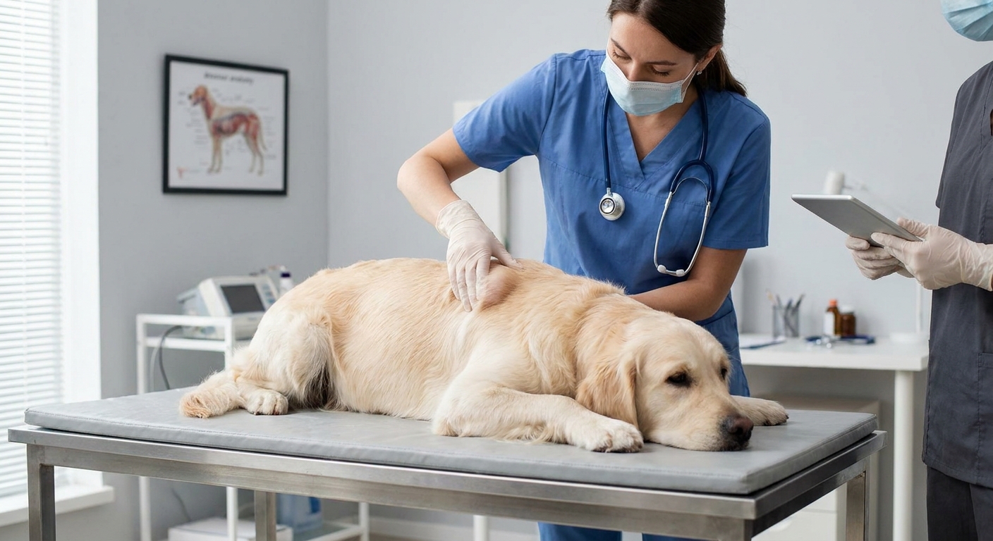
An infiltrative lipoma is a tumor made of fat cells, similar to a typical lipoma, but with one key difference: instead of staying neatly contained under the skin, it extends into nearby tissues, most often between and within muscle layers. Some can also involve fascia (the connective tissue around muscles), tendons, or even nearby nerves.
Because the cells themselves look benign under the microscope, infiltrative lipomas are not considered malignant cancers. The important caveat is that they can still cause real problems by:
- Invading muscle, making clean surgical removal difficult
- Limiting movement or causing discomfort depending on location
- Returning (recurring) after surgery if any infiltrating strands are left behind
Which dogs get them?
Infiltrative lipomas are most often diagnosed in middle-aged to older dogs. Some breeds are reported more commonly in veterinary literature (for example, Labrador Retrievers and Doberman Pinschers), but any dog can develop a fatty mass. Breed trends can vary between studies, so your dog’s lump still deserves an individualized workup.
Why it can feel like a simple lipoma
From the outside, an infiltrative lipoma may feel a lot like a classic fatty lump. It can be:
- Soft or doughy
- Slow-growing at first
- Not painful when touched
- Hard to define with your fingers if it blends into deeper tissues
The difference is that a superficial lipoma usually sits in the fatty layer just under the skin and tends to be more movable. An infiltrative lipoma may feel less “slippery” under your hand or seem tethered deeper, especially if it is threaded into muscle.
Still, finger-feel is not enough to diagnose any lump. Plenty of serious tumors can start out soft and innocent-seeming.
Infiltrative lipoma vs other lumps
Here is a puppy-scout rule that saves dogs: do not diagnose lumps by vibes. Vets separate look-alike lumps by doing a physical exam and sampling cells or tissue.
Infiltrative lipoma vs superficial lipoma
- Superficial lipoma: Benign fat tumor, typically under the skin, often very movable, usually well circumscribed.
- Infiltrative lipoma: Benign fat tumor that extends into muscle and connective tissue, can feel less movable, harder to remove fully, recurrence is more likely.
Infiltrative lipoma vs mast cell tumor
Mast cell tumors can be masters of disguise. They can look like almost anything: soft, firm, tiny, large, hairless, red, scabby, or totally normal.
- Mast cell tumor: A skin tumor that can be malignant. It may change size quickly, become red or itchy, ulcerate, or swell after being bumped or handled due to histamine release.
- Infiltrative lipoma: Usually slow and quiet on the skin surface, but invasive deeper in the body.
Because mast cell tumors can release histamine and cause local swelling, a lump that “puffs up” or changes day to day deserves prompt attention. This is also why repeatedly squeezing or poking a lump is a bad idea. You can trigger extra swelling and inflammation, and you can make your dog sore.
Infiltrative lipoma vs soft tissue sarcoma
Soft tissue sarcomas are a broad group of malignant tumors arising from connective tissues. Some grow slowly and feel deceptively similar to benign masses.
- Soft tissue sarcoma: Malignant, tends to invade locally and can recur. Some types can spread (metastasize) depending on grade.
- Infiltrative lipoma: Benign fat cells, but still infiltrative locally and prone to recurrence if not fully removed.
The important overlap: both can infiltrate. That is why imaging and tissue diagnosis matter, especially for deeper, fixed, or fast-growing masses.

Where infiltrative lipomas show up
These tumors can occur in different areas, but they are often discussed in relation to:
- Limbs and shoulder region
- Chest wall
- Neck or back
- Areas with substantial muscle layers
A lump that seems deep, broad, or difficult to “pinch up” with the skin can be a clue that a mass involves deeper tissue, although that still does not tell you what type it is.
How vets diagnose an infiltrative lipoma
Diagnosis is usually a stepwise puzzle, and it is the safest path for your dog. This article is for education, not a substitute for veterinary diagnosis.
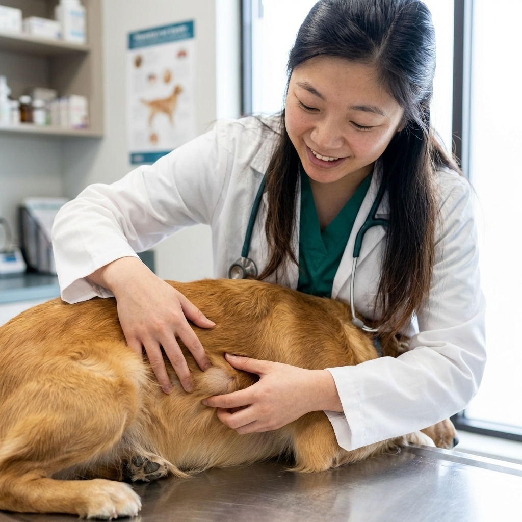
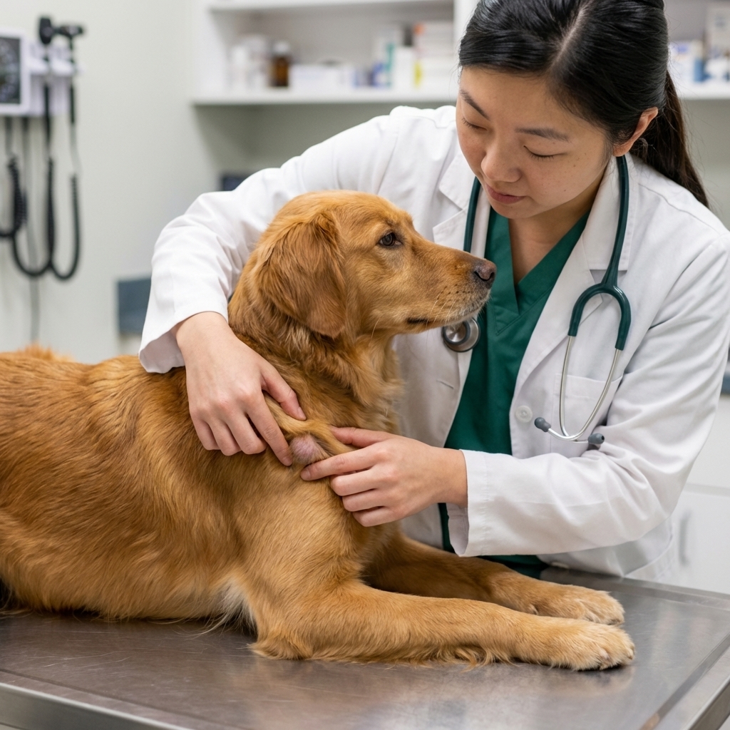
Physical exam and history
Expect questions like: How long has it been there? Is it changing quickly? Does your dog lick at it? Any limping or stiffness?
Fine needle aspirate (FNA)
An FNA uses a small needle to collect cells. If the sample shows mostly fat cells, it can suggest a lipoma, but it cannot always confirm whether the mass is infiltrative. Fat can be frustratingly non-diagnostic on cytology, especially with deep masses or mixed tissues.
Imaging
For suspected infiltrative lipomas, imaging helps map how far the mass extends and what structures are involved.
- Ultrasound can help assess depth and relationships, and it may guide sampling, but it does not reliably sort infiltrative lipomas from other infiltrative masses on its own.
- CT or MRI are often most useful for surgical planning because they can show how a mass tracks between muscle layers and around important structures.
Biopsy and histopathology
A biopsy, followed by lab evaluation of tissue, is the most definitive way to identify what a mass is. Histopathology can also help rule out malignant tumors that can mimic benign ones.
Why removal is wider and recurrence is more likely
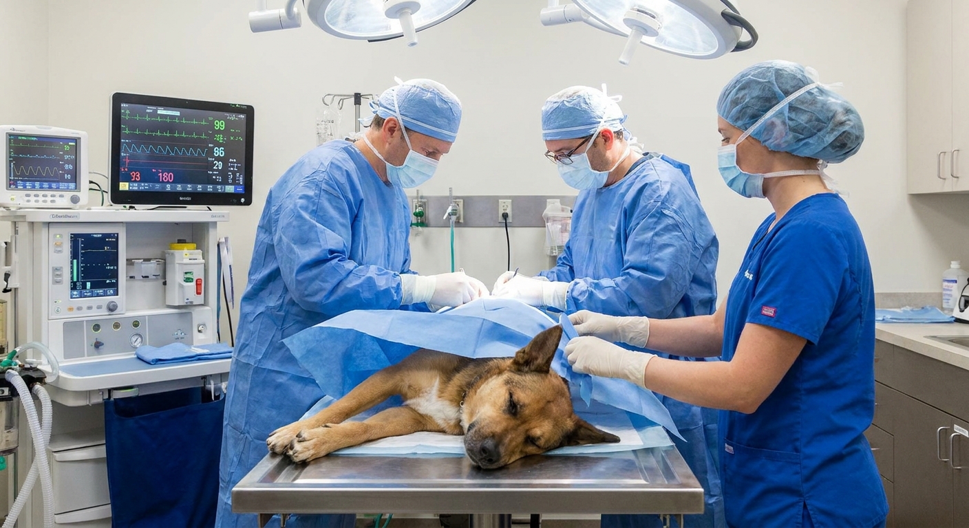
With a superficial lipoma, a surgeon can often “shell out” the mass because it is more neatly separated from surrounding tissue.
Infiltrative lipomas are different. They can send fat between muscle bundles like roots through grass. That means:
- Wider surgical margins may be recommended to reduce the chance of leaving behind infiltrating cells.
- Surgery can be more complex depending on proximity to nerves, blood vessels, or joints.
- Recurrence is more likely if microscopic extensions are left in place, even when the main mass is removed.
If your vet talks about “margins,” they mean the rim of normal-looking tissue taken around the lump. Margins can be evaluated by a pathologist. “Clean” or “complete” margins suggest the mass was likely removed fully. “Dirty” or “incomplete” margins mean tumor cells extend to the edge of the sample, so regrowth is more likely.
In some cases, your veterinarian may discuss additional strategies like referral to a surgical specialist or considering radiation therapy when complete excision is difficult. The right plan depends on the tumor location, size, your dog’s comfort and mobility, and what diagnostics show.
What owners can watch for at home
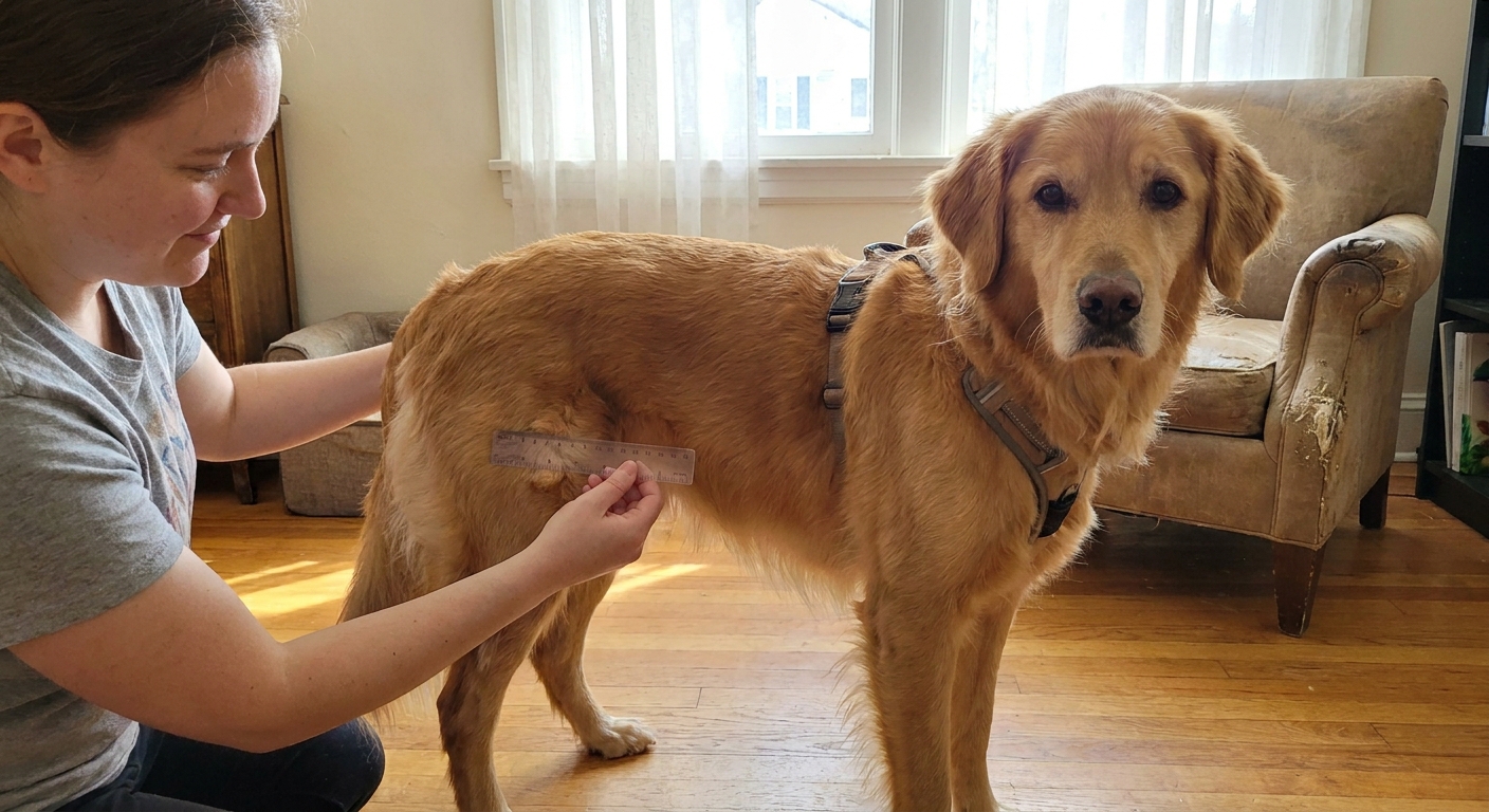
I love a good home “sniff and check,” as long as it stays gentle and does not replace a vet visit. If you find a lump, note:
- Location (front leg, ribcage, belly, etc.)
- Size (use a ruler or compare to a coin, then write it down)
- Texture (soft, firm, rubbery)
- Mobility (does it slide under the skin or feel fixed?)
- Skin changes (redness, scabbing, hair loss, sores)
- Behavior changes (licking, sensitivity, limping)
Taking clear photos every couple of weeks can help you and your vet track changes. Do not squeeze hard or keep poking a lump “to see if it changed.” Aside from irritating the tissue, some tumors (especially mast cell tumors) can swell with manipulation due to histamine release.
Lump changes to take in promptly
Any new lump is worth scheduling, but some changes should move you up the calendar. Contact your veterinarian promptly if a lump:
- Appears suddenly or grows quickly over days to weeks
- Changes shape or becomes more firmly attached
- Becomes red, swollen, bruised-looking, or warm
- Ulcerates (opens, bleeds, or oozes)
- Seems painful, or your dog resists being touched
- Triggers licking, chewing, or scratching
- Is near the mouth, eyes, anus, genitals, or on a foot (these areas can be higher stakes)
- Is associated with limping, weakness, or reduced range of motion
If you remember only one puppy-scout note: “It’s probably a lipoma” is not a diagnosis. A quick needle sample can save a lot of worry or catch something serious early.
Treatment and outlook
The outlook for infiltrative lipomas is often good in the big picture because they are benign and do not behave like metastatic cancers. The practical challenge is local control.
Common treatment options
- Surgical removal, often with a plan for wider margins than a superficial lipoma
- Specialty surgery when the mass is deep, extensive, or near important structures
- Radiation therapy may be considered in selected cases when complete removal is not possible or recurrence would be especially problematic
- Follow-up monitoring for recurrence, especially in the months and years after removal
What to expect after treatment
Recurrence, when it happens, can show up months or even years later. Your vet may recommend rechecks on a schedule (often more frequent in the first year), and they may suggest you continue your at-home measuring and photo routine. If your dog had advanced imaging for surgical planning, your vet may use those results to guide what “watching for recurrence” looks like for that exact location.
Ask your veterinarian what they saw during surgery, whether margins were discussed, what the pathology report showed, and what the follow-up plan is for your dog’s specific case.
FAQ
Is an infiltrative lipoma cancer?
It is considered benign, meaning it is not malignant. But it can still be locally invasive, which is why it can require more advanced planning and follow-up than a typical lipoma.
Can an infiltrative lipoma turn into cancer?
In most cases, benign lipomas are not thought to transform into malignant tumors. The bigger issue is that different tumors can look similar from the outside. Rare malignant fat tumors (such as liposarcomas) do exist and typically arise on their own rather than from a known benign lipoma. Sampling and, when needed, biopsy are important for a true diagnosis.
How can my vet tell it is infiltrative?
Imaging (especially CT or MRI) can show the extent into muscle, and histopathology from a biopsy or removed tissue can confirm the tumor type.
Should I remove a lump if my dog seems fine?
That is a decision to make with your veterinarian. Some lumps can be monitored. Others are best removed early, especially if they are growing, deep, interfering with movement, or located where surgery will be more difficult later.
The puppy-scout takeaway
Infiltrative lipomas are the “looks simple, behaves complicated” kind of lump. They can feel like a regular fatty tumor, but because they grow into muscle, they often need wider removal and careful follow-up for recurrence.
If your dog has a new lump, or an old lump that is changing, your best next step is wonderfully unglamorous: book the vet visit and get the sample. Your dog will forgive you for the appointment. Especially if there are treats.