A curious nose. A very waggy welcome.
Sometimes a “weird spot” is just a scab that heals. Sometimes it is a stubborn sore, an odd nail problem, or a mouth bump you only notice when your dog yawns. One diagnosis vets take seriously in all of those places is squamous cell carcinoma, often shortened to SCC.
SCC is a cancer that starts in squamous cells, which are common surface lining cells in the skin and in mucous membranes such as the mouth. Where SCC shows up matters a lot because how it behaves, how it gets biopsied, and how it is staged can change by location.

What is squamous cell carcinoma in dogs?
Squamous cell carcinoma is a malignant tumor of squamous epithelium. In plain terms, it is cancer that develops from the cells that make up parts of the outer skin and the lining of certain body surfaces.
In dogs, SCC often behaves as a locally invasive cancer. That means it can dig into nearby tissues and destroy them. Depending on where it forms, it may or may not spread to distant sites, but metastasis can occur, especially in more aggressive cases or later stages.
Where SCC is commonly found
- Skin (often on sun-exposed areas, especially thin-haired or lightly pigmented spots)
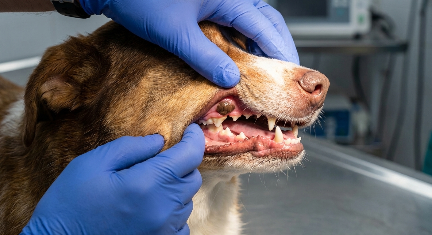
- Mouth (oral SCC)
- Nail bed (digital SCC, around a toenail)
SCC vs melanoma: what is different?
SCC and melanoma can show up in some of the same areas on a dog, like toes and mouths, but they come from different cell types and often look and act different.
Cell of origin
- SCC starts in squamous epithelial cells, the “lining” type cells.
- Melanoma starts in melanocytes, the pigment-making cells.
How they tend to look
- SCC commonly appears as a crusty sore, ulcer, wart-like growth, thickened plaque, or a non-healing lesion. Pigment is not the main feature, although lesions can look red, pink, gray, or mixed.
- Melanoma is often darkly pigmented, but not always. Some melanomas are pink or minimally pigmented, which is why appearance alone cannot rule it out.
How they tend to behave
Both cancers can be serious. As a broad pattern, SCC is often described as locally invasive first, with metastasis risk that depends on location and stage. Melanoma, especially in the mouth, is often treated as having a higher metastatic potential. But the only safe rule is this: you cannot accurately predict behavior from a photo or a guess. Diagnosis and staging matter.
If a vet is choosing between SCC and melanoma on the list, it usually means the next step is the same: get a sample and let pathology name it.
SCC on skin (often sun-exposed)
Cutaneous SCC can develop on many parts of the body. One well-known pattern is SCC that develops on areas that get a lot of sun exposure and have less natural protection from pigment or thick fur.
That said, SCC can also show up in less sun-exposed spots. The UV association is considered strongest for certain cutaneous forms and locations, so your veterinarian will still keep SCC on the list even when the “sun story” is not obvious.
Common locations
- Ear tips and pinna (ear flap)
- Nose and nasal planum
- Eyelids
- Belly and inner thighs in dogs who sunbathe belly-up
What it can look like
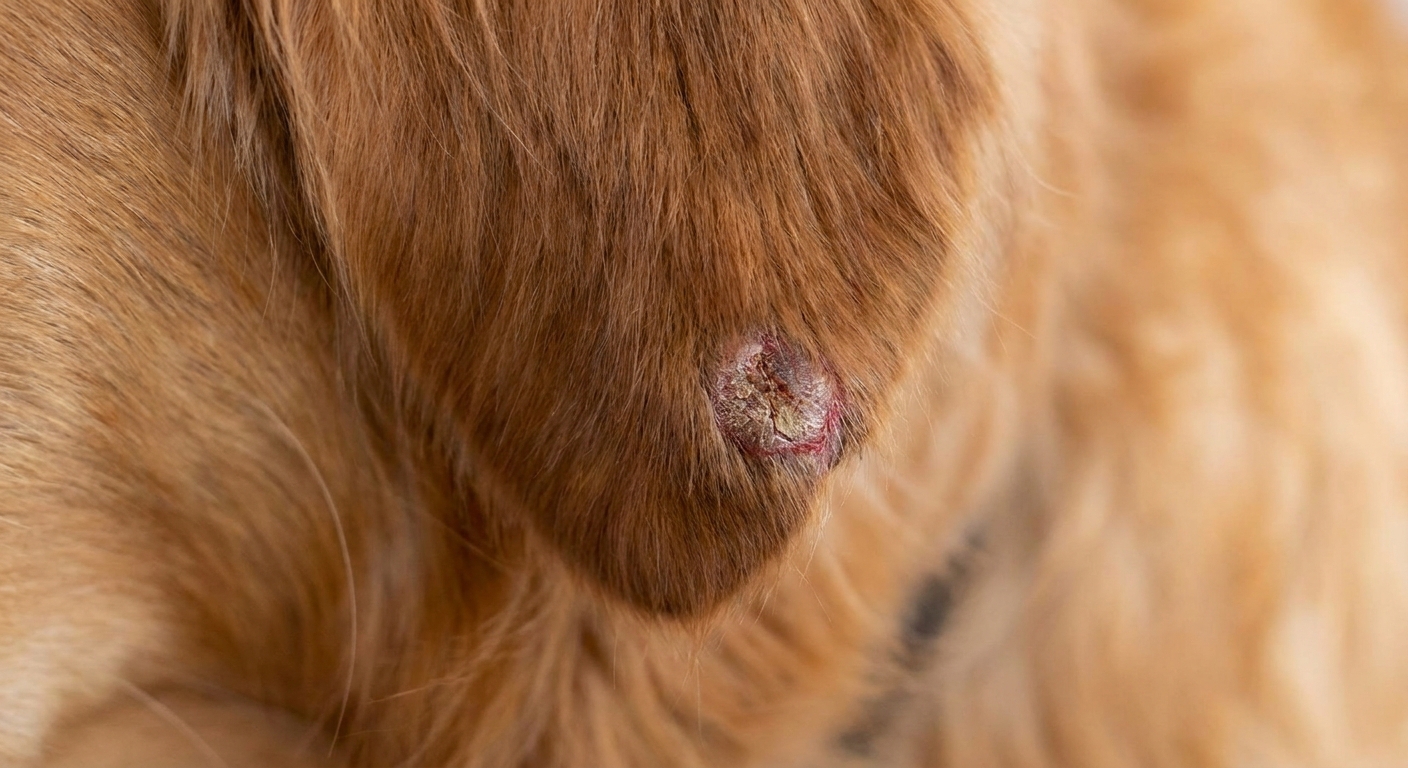
- A scaly, crusty patch that keeps coming back
- A non-healing sore or ulcer
- A raised, irregular growth that bleeds easily
- Thickened, rough, or “cauliflower-like” skin texture
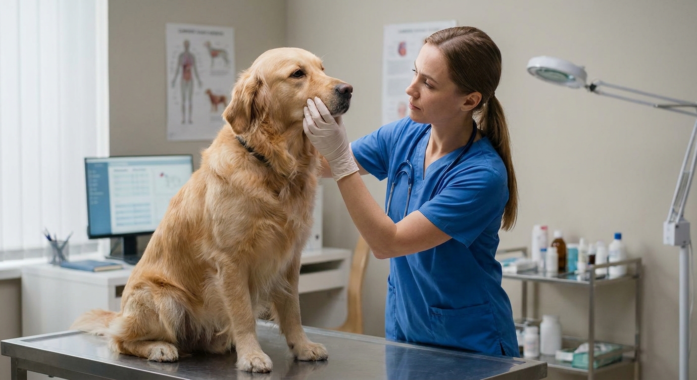
Biopsy and diagnosis for skin SCC
Skin lesions are often sampled in a few ways:
- Fine needle aspirate (FNA): a small needle pulls out cells for review. This can be helpful, but SCC does not always give a clean, definitive answer on FNA, especially if the sample is inflamed or crusted.
- Biopsy: a small piece is removed and sent to a pathologist. For SCC, this is often what confirms the diagnosis.
- Excisional biopsy: the vet removes the entire mass (if feasible) and sends it in. This can diagnose and treat in one step when the lesion is small and in a removable spot.
Pathology reports often mention things like degree of differentiation (how much the cancer cells resemble normal cells) and whether the sample suggests a more aggressive pattern.
What staging means for skin SCC
Staging is the process of checking how extensive the cancer is. For skin SCC, staging is often focused on:
- Measuring the tumor and evaluating depth and local invasion
- Checking nearby lymph nodes for enlargement and sampling them if suspicious
- Looking for spread in higher risk cases, which may include chest imaging and other tests based on the individual dog
The goal is to guide treatment planning, not to “assign a label” for its own sake. A small, removable SCC with no evidence of spread is very different from a large lesion that has invaded deeper tissues.
Oral squamous cell carcinoma (mouth SCC)
Oral SCC is among the more common malignant oral tumors in dogs. In many references, oral melanoma is cited as the most common malignant oral tumor, with SCC commonly next. Either way, the mouth has lots of important real estate packed into a small space, so even a tumor that prefers to stay local can cause big problems.
Signs you might notice at home
- Bad breath that seems new or unusually strong
- Drooling, sometimes with blood
- Reluctance to chew or dropping food
- Pawing at the mouth
- Facial swelling
- Loose teeth that do not match your dog’s age or dental history
How oral SCC differs from many mouth melanomas
Both can be aggressive in the mouth, but they are not the same disease. Oral SCC often causes significant local invasion into gums, bone, and surrounding tissues. Oral melanoma is often treated as having a notable risk of metastasis. Either way, the mouth is not a wait-and-see location.
Biopsy and diagnosis for oral SCC
Oral tumors usually need a tissue biopsy for a confident diagnosis. Because the mouth bleeds easily and the sample needs to be deep enough, this is often done with sedation or anesthesia.
A biopsy can be:
- Incisional biopsy: taking a piece of the mass to get a diagnosis before planning a larger surgery.
- Excisional biopsy: removing the whole visible mass. In the mouth, this is not always possible or recommended until imaging and surgical planning are done.
In some cases, the care team may recommend imaging before or soon after biopsy, especially if they suspect bone involvement. Common examples include dental radiographs and CT, which help assess jaw involvement and guide surgical margins.
What staging means for oral SCC
Staging for oral SCC often focuses on three big questions:
- How big is it and where exactly is it? Size and location affect surgical options.
- Has it invaded bone? Oral SCC can invade the jaw. Imaging helps evaluate this and plan margins.
- Has it spread? Vets may assess regional lymph nodes and may recommend chest imaging, because staging helps decide whether local treatment alone is enough.
If your vet talks about “staging,” it usually means they are building a map before choosing the route.
Nail bed squamous cell carcinoma (toe and nail SCC)
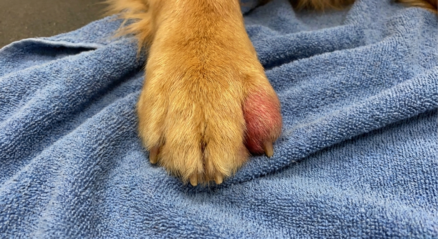
Nail bed SCC is one of the sneakiest forms because it can look like an everyday nail injury for a long time. Dogs break nails, lick sore feet, and get infections. SCC can wear that same costume.
Some risk patterns have been reported for digital SCC, including higher occurrence in certain large breeds and in darker-coated dogs. But no pattern is strong enough to safely ignore a stubborn single-toe problem in any dog.
Clues that a “nail problem” might be more than a nail problem
- A nail that keeps breaking or falls off repeatedly
- Swelling of one toe that does not resolve
- Persistent licking of one foot
- Drainage, bleeding, or a chronic “infected” look around the nail
- Lameness focused on a single toe
- A firm mass at the base of the nail
Biopsy and diagnosis for nail bed SCC
For nail bed tumors, superficial samples can miss the real issue. Diagnosis commonly requires a tissue biopsy, and because toes are sensitive, that often means sedation or anesthesia.
Depending on how the toe looks, your veterinarian may recommend:
- Biopsy of the nail bed mass
- Toe radiographs (X-rays) to look for bone changes
- Amputation of the affected digit with the removed tissue sent to pathology, which can be both diagnostic and therapeutic in many cases
What staging means for nail bed SCC
Staging for digital SCC may include:
- Evaluating the toe bone (local invasion is common, and X-rays can help assess it)
- Checking regional lymph nodes (feeling them, imaging them, and sometimes sampling them)
- Screening for spread in selected cases, which may include chest imaging and additional testing guided by your vet
One reason staging matters here is that treatment often involves local control (like removing the affected digit) and your care team wants to know if anything else needs attention beyond the toe.
How vets confirm SCC: what a biopsy report can tell you
A biopsy is not just a “yes or no” for cancer. Pathology can provide details that shape the plan.
Common biopsy terms you might see
- Diagnosis: squamous cell carcinoma (or something else)
- Margins: whether cancer cells extend to the edge of the removed tissue, suggesting tumor may have been left behind
- Differentiation (and sometimes grade): how abnormal the cells look and how they are organized, which can relate to expected behavior
- Invasion: whether there is evidence of deeper infiltration into surrounding tissue
If margins are not clean, that does not automatically mean disaster. It means your vet may discuss next steps such as a wider surgery, radiation, or other options depending on location.
Treatment basics, by location
Treatment choices depend on location, size, invasion, and what staging shows. Your veterinarian may also suggest referral to a veterinary oncologist, surgeon, or dental specialist for oral cases.
Skin SCC
- Surgery is often the main treatment when the lesion can be removed with adequate margins.
- Radiation may be considered when surgery cannot get clean margins or the location makes wide removal difficult.
- Sun management becomes part of long-term prevention and management for susceptible areas.
Oral SCC
- Surgery can be more complex because of local invasion and the need for functional margins.
- Radiation is sometimes used as a primary or additional therapy depending on case details.
- Pain control and nutrition support are not “extras” in mouth cancers. They are core quality-of-life pieces.
Nail bed SCC
- Digit amputation is commonly recommended because it can remove the local tumor and address bone involvement.
- Lymph node evaluation and additional therapy may be discussed depending on staging results.
What about chemotherapy or other systemic treatment?
For many SCC cases, treatment is focused on local control (surgery, radiation, or both). If there is evidence of spread, or if local control is not possible, your vet or oncologist may discuss systemic options such as chemotherapy or other medications as part of the plan.
Prognosis depends on location and timing
I wish SCC came with one neat, universal forecast. It does not. A small SCC on a sun-exposed ear tip that can be fully removed is a different situation than an oral SCC that has invaded jawbone, and both are different from a nail bed SCC that has been quietly simmering for months.
What tends to help across the board is catching it earlier, when the tumor is smaller and the treatment menu is wider.
When to call your vet quickly
I know, I know. Dogs get bumps and scrapes like it is their hobby. Still, these signs deserve a prompt call and an appointment:
- A sore on the ear, nose, eyelid, or belly that does not improve within about 1 to 2 weeks
- A skin lesion that bleeds easily, crusts repeatedly, or enlarges
- Bad breath plus drooling, bleeding, or trouble eating
- A single swollen toe, recurring broken nail, or chronic “infected” nail bed
- Any mouth mass you can see, especially if it seems to grow
If your dog seems painful, stops eating, or is limping significantly, treat that as urgent.
Can SCC be prevented?
Not all SCC is preventable, but risk reduction is a real thing, especially for sun-exposed skin.
Helpful prevention habits
- Limit intense sun exposure during peak hours for dogs who love sunbathing.
- Provide shade outdoors and encourage breaks from direct sunlight.
- Monitor high-risk spots like ear tips, nose, and thin-haired bellies.
- Do regular “nose to toes” checks so you notice changes early.
If you are considering sunscreen for a dog with sensitive, lightly pigmented skin, ask your veterinarian what is safe. Many human sunscreens contain ingredients that are not dog-friendly if licked. When sun protection is needed, physical barriers and shade are often the simplest, safest place to start.
Quick questions I hear a lot
Does SCC always spread?
No. SCC is often most destructive where it starts. But it can spread, and the risk depends on factors like location, size, invasion, and what staging finds.
Can an FNA diagnose SCC?
Sometimes, but SCC can be tricky on cytology alone. For many suspected SCC cases, a biopsy is what provides a clear answer.
Why does location matter so much?
Because “SCC” is a diagnosis, not a map. Skin SCC on an ear tip, oral SCC near the jawbone, and nail bed SCC at a toe can each have different surgical options, different staging needs, and different quality-of-life considerations.
What should I bring to the appointment?
A timeline helps. Note when you first saw the lesion, whether it changes size, bleeds, crusts, or seems painful. If it is on a toe or in the mouth, a short video of limping or chewing trouble can also be useful.
The takeaway
Squamous cell carcinoma in dogs is a cancer of surface lining cells that commonly appears on skin (often sun-exposed areas), in the mouth, or at the nail bed. It is often locally invasive, and it can sometimes spread. The big power tools here are biopsy to name what it is and staging to understand how far it goes in that specific location.
If you are staring at a stubborn sore, a mysterious mouth bump, or a toe that just will not heal, the best next step is not more guessing. It is a vet visit and a plan.