
Soft Tissue Sarcoma in Dogs: When a Movable Lump Matters

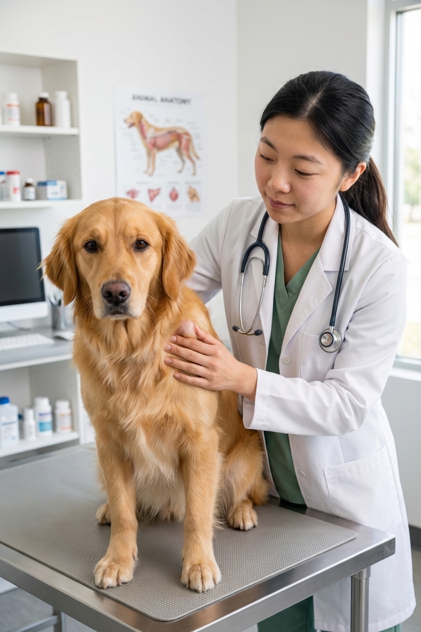
If you have ever found a lump on your dog and thought, “It moves around, so it must be a lipoma,” you are in very good company. I hear this all the time in clinic. And to be fair, many movable, soft lumps are benign fatty tumors.
But here is the important truth: some cancers, including soft tissue sarcomas, can feel deceptively “friendly” at the surface, especially early on. They may seem smooth and mobile. Underneath, tumor cells can still be extending into nearby tissue where you cannot feel them.
Let’s walk through what soft tissue sarcoma is, how it differs from a lipoma and from mast cell tumors, what biopsy and staging mean in everyday language, and what treatment often looks like.
Lipoma vs soft tissue sarcoma
How a typical lipoma feels
A lipoma is a benign tumor made of fat cells. Typical lipomas are:
- Soft or doughy
- Easy to move under the skin
- Slow growing
- Not painful
Most lipomas do not invade surrounding tissue the way cancers do. One important exception is an infiltrative (intramuscular) lipoma, which can extend between muscle layers and be harder to fully remove. Those behave differently than the common “simple” lipoma, and your vet may recommend imaging or a different surgical plan if one is suspected.
How a soft tissue sarcoma can feel
Soft tissue sarcoma (STS) is a broad category of cancers that arise from connective tissues such as fibrous tissue, muscle, nerve sheath tissue, and related structures. Many STS lumps are:
- Firm to rubbery, but not always
- Sometimes movable, especially early when they are more superficial
- Slow to moderately growing, with some exceptions
- Often painless
Here is the key difference: STS commonly grows with microscopic “fingers” extending into surrounding tissue. So even if the lump seems to slide under your fingers, the tumor can still be anchored deeper where you cannot feel it.

What is a soft tissue sarcoma?
Soft tissue sarcoma is not one single cancer type. In dogs, it is an umbrella term that often includes tumors such as:
- Peripheral nerve sheath tumors
- Fibrosarcoma
- Myxosarcoma
- Perivascular wall tumors
- Other related soft tissue tumors
From an owner perspective, the most important pattern to understand is this:
- Local behavior: STS tends to be very good at coming back in the same place if it is not removed with wide margins.
- Spread (metastasis): Many STS have a lower metastatic rate than some other cancers, but risk varies by tumor grade and type. When they do spread, it is most commonly to the lungs. Lymph nodes or other organs are less common, but possible, especially with higher-grade tumors or certain subtypes.
Why a movable lump can be serious
When a lump sits in the fatty layer under the skin, it can feel mobile even if it is malignant. What makes sarcomas tricky is that they can:
- Look like a harmless bump for weeks or months
- Feel smooth on the surface
- Be covered by normal-looking skin
- Still send microscopic cells into nearby fascia and muscle
That infiltrative growth is why your veterinarian may recommend a more proactive plan for a suspected sarcoma than for a typical lipoma, even if the lump “seems easy.”
First step: FNA vs biopsy
Fine needle aspirate (FNA)
An FNA uses a small needle to collect cells from the lump. It is quick and usually does not require sedation.
Pros: inexpensive, fast, minimally invasive.
Limitations for sarcoma: STS can be difficult to diagnose by cytology alone because the cells may not exfoliate well. Results sometimes come back as “spindle cells,” “mesenchymal cells,” or “inconclusive.” That does not mean it is benign. It often means we need more information.
Biopsy
A biopsy takes a larger tissue sample so a pathologist can evaluate the architecture of the tumor, not just individual cells.
- Incisional biopsy: a piece is taken, the tumor stays in place for now.
- Excisional biopsy: the lump is removed entirely and submitted.
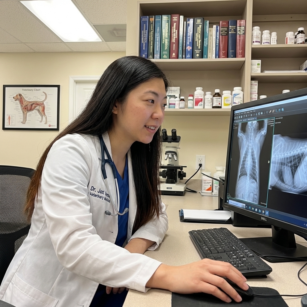
Definitive diagnosis and grading require histopathology. Cytology can sometimes suggest sarcoma, but it generally cannot provide a reliable grade the way a tissue sample can.
For suspected STS, many veterinarians prefer an approach that supports a future “clean margin” surgery. That may mean an incisional biopsy first, imaging first, or referral to a surgeon, depending on location and size.
If your vet says, “Let’s sample this before we remove it,” it is usually to avoid a scenario where a sarcoma is removed with narrow margins and then is more likely to recur and be harder to control after that first surgery.
Staging basics
When owners hear “staging,” it can feel scary. Staging is simply a way to answer two practical questions:
- How aggressive is this tumor likely to be? (grade)
- Has it spread anywhere else? (metastasis check)
Grade: low, intermediate, or high
STS is often assigned a tumor grade by the pathologist based on things like how abnormal the cells look, how quickly they are dividing, and how much tissue death is present within the tumor. In general:
- Low-grade tumors tend to spread less often but can still recur locally if not removed widely.
- High-grade tumors have a higher risk of metastasis and may drive more intensive treatment recommendations.
Common staging tests
- Chest X-rays or a CT scan to look for lung spread
- Bloodwork to assess overall health and anesthesia readiness
- Sampling lymph nodes if they are enlarged or if the tumor type or location raises concern
- Imaging the mass (often ultrasound, CT, or MRI) to see how deep it goes and to help plan surgery
Treatment basics
Surgery: the cornerstone
For many soft tissue sarcomas, surgery is the main treatment. The goal is not just to remove the visible lump, but to remove a “buffer zone” of normal tissue around it. This is called getting margins.
In plain language:
- Clean margins means no tumor cells are seen at the cut edge.
- Close margins means tumor cells are near the cut edge.
- Dirty (incomplete) margins means tumor cells extend to the cut edge.
With STS, margins matter because those microscopic tumor extensions can be left behind even when the lump looks completely removed to the naked eye.
Radiation therapy
Radiation is often recommended when:
- The tumor is in a spot where wide margins are difficult (leg, face, near joints)
- Surgical margins are close or incomplete on the pathology report
- The tumor is likely to recur and a second surgery would be tough
Radiation can be used after surgery to control microscopic disease, or in some cases before surgery to shrink or better define the tumor field.
Chemotherapy
Chemotherapy is not automatically part of every STS plan. It is more likely to be discussed when:
- The tumor is high grade
- There is evidence of metastasis
- The specific sarcoma subtype has higher spread risk
Your veterinary oncologist will tailor recommendations to the grade, location, and your dog’s overall health.
When referral matters
Because the first surgery is often the best chance for long-term local control, referral is worth discussing if:
- The mass is on a limb, near a joint, or in a location where margins will be challenging
- The mass is large, rapidly growing, or fixed to deeper tissue
- Advanced imaging (CT or MRI) would change the plan
- Radiation therapy may be needed and a facility is not local
A board-certified surgeon or surgical oncologist can help plan the incision, margins, and reconstruction. A veterinary oncologist can help integrate surgery, radiation, and chemotherapy when appropriate.
Mast cell tumor vs sarcoma
We cover mast cell tumors (MCT) in a separate article, and that comparison is helpful because mast cell tumors come with their own special urgency and precautions.
Here are the practical differences owners should know:
Mast cell tumors
- Can change size quickly, sometimes swelling and shrinking
- May cause itchiness, redness, hives, GI upset, or ulceration due to histamine release
- Often prompt vets to recommend quicker evaluation and careful handling
Soft tissue sarcomas
- Often grow slowly and quietly, which can falsely reassure owners
- Usually do not cause systemic histamine-type reactions
- Create a “surgical planning” urgency because first surgery is the best chance for long-term local control
So while MCT urgency often relates to biologic behavior and mediator release, STS urgency is frequently about not missing the window for a well-planned, wide-margin removal.
What you can track at home
Before your visit (or between rechecks), a little tracking can be surprisingly helpful:
- Measure it with a ruler in two directions (for example, length and width in centimeters).
- Take a photo with the ruler in the picture for scale.
- Note the location (for example, “right chest wall, two inches behind the elbow”).
- Watch the trend: is it stable, slowly growing, or changing quickly?
This does not replace sampling, but it helps your vet understand growth rate and plan the next steps.
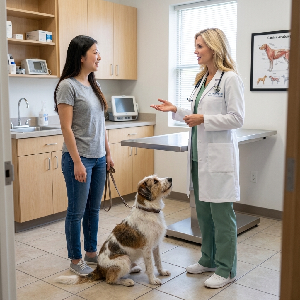
When to call your vet sooner
If you find a new lump, I always recommend scheduling an exam. And if any of these apply, move it up on your calendar:
- The lump is growing over days to weeks
- It is firm, irregular, or seems attached deeper than the skin
- It is on a limb or near a joint where surgery margins are tougher
- The skin over it looks bruised, ulcerated, or inflamed
- Your dog seems painful, is licking it, or it interferes with movement
A helpful rule of thumb many vets use: any lump that is growing, any lump larger than about 1 inch (2.5 cm), or any lump present for a month deserves a veterinary check and a discussion about sampling.
What to ask at your appointment
These questions can help you leave the clinic with a clear plan:
- Can we do an FNA today, and if results are unclear, what is our next step?
- If you suspect sarcoma, should we do a biopsy before removal?
- Would imaging like CT or ultrasound change the surgical plan?
- If this is removed, will it be submitted for histopathology automatically?
- If margins come back close or incomplete, what are the next options: re-excision, radiation, or referral?
Bottom line
A movable lump is not a diagnosis. Many are lipomas, and I love when we get that good news. But soft tissue sarcomas can start out subtle and still be serious because of how they infiltrate surrounding tissue.
Prognosis varies widely. Many low-grade sarcomas do very well with good local control, especially when removed with wide margins (and radiation when needed). Higher-grade sarcomas carry more metastatic risk and may require a more layered plan. Either way, early sampling and smart surgical planning usually put you in the best possible position.
Quick reminder: This article is educational and not a substitute for veterinary care. If you find a new lump or an old lump that is changing, schedule an exam and ask about FNA or biopsy.