
Found a new lump on your dog? Compare lipoma vs mast cell tumor clues, know the warning signs, and learn why an FNA or biopsy is the only way to confirm what...
Article
•
Designer Mixes
Mast Cell Tumors in Dogs: Grades, Treatment, and Life Expectancy

Shari Shidate
Designer Mixes contributor
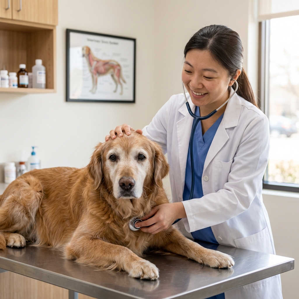
Mast cell tumors (MCTs) are one of the most common malignant skin tumors in dogs. If you have found a new lump on your dog, you are not overreacting by wanting it checked quickly. Mast cell tumors can look like almost anything: a small, soft bump, a firm nodule, a scab that will not heal, or a “bug bite” that changes size from day to day.
As a veterinary assistant in Frisco, Texas, I have seen how scary the waiting can feel. The good news is that many mast cell tumors are very treatable, especially when we catch them early and get the right staging and a clear treatment plan.

What mast cell tumors are (and why they can act unpredictably)
Mast cells are normal immune cells that live in tissues throughout the body. They release chemicals like histamine as part of allergic and inflammatory responses. When mast cells become cancerous, they can form tumors most often in the skin, but also sometimes in places like the spleen, liver, intestines, or bone marrow.
One reason MCTs can be tricky is that the tumor can “degranulate,” meaning it releases histamine and other inflammatory chemicals. This can cause:
- Redness, swelling, itching, and bruising around the mass
- The lump to get larger or smaller over hours or days
- Stomach upset, poor appetite, vomiting, or dark stools in some dogs
- Rarely, more serious allergic-type reactions during handling or surgery
If you ever notice your dog has a lump that suddenly swells, becomes red, or seems painful, contact your veterinarian as soon as you can.
Common signs owners notice
Mast cell tumors are sometimes called “the great pretenders” because they can mimic many other skin growths. Signs that warrant a vet visit include:
- A new lump anywhere on the body, especially one that changes size
- A lump that looks inflamed or gets puffy after your dog scratches or you touch it
- A skin mass that ulcerates, bleeds, or does not heal
- Multiple lumps developing over time
- Unexplained vomiting, diarrhea, or black, tarry stool (possible GI ulceration)
- Lethargy or decreased appetite
Not every lump is cancer, but every lump deserves a plan. Early evaluation gives you the most options.
One reassuring note: multiple separate skin MCTs do not always mean the cancer has spread. Some dogs develop more than one primary mast cell tumor over time. They still deserve a careful workup, but “more than one” is not automatically the same thing as metastatic disease.
Risk factors and breeds
Any dog can develop an MCT, but we do see them more often in certain breeds. Commonly cited higher-risk breeds include Boxers, Boston Terriers, Bulldogs, Pugs, Labrador Retrievers, and Golden Retrievers.
This does not mean mixed breeds are “safe,” or that a low-risk breed cannot get one. It just helps explain why your vet may take certain lumps more seriously in some dogs.
Diagnosis: what to expect at the vet
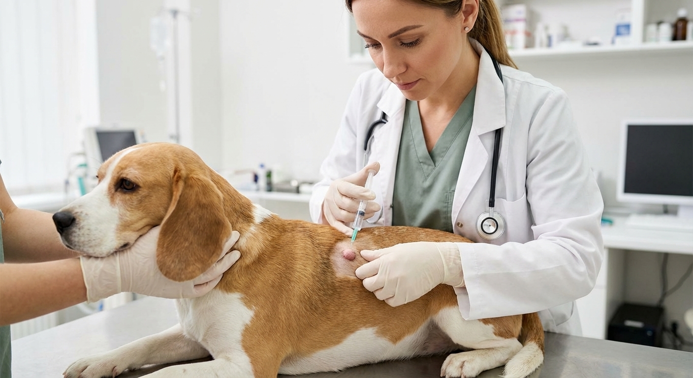
Fine needle aspirate (FNA)
In many cases, the first step is an FNA, where a small needle collects cells from the lump. It is quick and often does not require sedation. Mast cells are frequently identifiable on cytology, so this test can provide fast answers.
Biopsy and histopathology
If the mass is removed or if cytology is unclear, your veterinarian may recommend a biopsy. Histopathology is how we determine tumor grade and whether the surgical margins are clean (meaning no tumor cells are seen at the edges of the removed tissue).
Staging tests (checking for spread)
Your dog may also need staging, depending on the tumor’s appearance, location, grade, and other risk factors. Staging can include:
- Bloodwork and urinalysis
- Lymph node aspirates (even if the node feels normal)
- Abdominal ultrasound (especially for higher-risk cases)
- Thoracic imaging in some cases (often more for overall cancer screening, anesthesia planning, or high-risk disease than because the lungs are the most common MCT target)
- Spleen and liver aspirates in select situations, most often when ultrasound findings are abnormal or in higher-risk or high-grade cases
Staging is not about “looking for bad news.” It is about choosing the treatment that fits your dog’s actual risk.
Staging: the quick overview
You may hear your veterinarian mention “stage,” which is different from “grade.” Grade describes how the cells look under the microscope. Stage describes how far the disease has spread in the body.
There are a few staging systems used in veterinary medicine, but a simple way to think about it is this:
- Lower stage: a single skin tumor with no evidence of spread to lymph nodes or internal organs
- Higher stage: involvement of lymph nodes, multiple tumors with concerning features, or spread to internal organs
If nearby lymph nodes are involved, it often changes the treatment plan and follow-up schedule, even if the original skin tumor was removed.
Mast cell tumor grades: what they mean
Grade describes how aggressive the tumor cells look under the microscope. Higher grade generally means a higher chance of regrowth or spread, but other factors matter too.
Patnaik grading system (Grade I, II, III)
- Grade I: Typically low grade and often cured with complete surgery.
- Grade II: Intermediate behavior, can be variable. Some behave like low grade tumors, some act more aggressively.
- Grade III: High grade, more likely to spread and recur.
Kiupel system (Low grade vs high grade)
Many labs now use the Kiupel system because it is more consistent:
- Low grade: Usually slower growing and more likely to be controlled with surgery.
- High grade: More aggressive with higher metastatic potential.
Beyond grade: other “big deal” predictors
Your veterinarian may discuss additional factors that help predict behavior:
- Margins: Whether the tumor was fully removed.
- Mitotic index: How quickly cells appear to be dividing.
- Location: Some locations (especially mucocutaneous areas like lips and mouth, and areas like the groin and perineum) can be harder to remove and may carry higher risk.
- Lymph node involvement: Spread to nearby nodes changes treatment planning.
- c-KIT mutations and KIT staining pattern: Can influence prognosis and use of targeted medications.
Treatment options
There is no one-size-fits-all plan for mast cell tumors. The best approach depends on grade, location, staging results, and what is realistic for your family.
1) Surgery
For most skin MCTs, surgery is the first choice when possible. The goal is to remove the tumor with an adequate margin of normal tissue around it. In some locations, wide margins are hard to achieve, so your veterinarian may refer you to a surgeon or recommend additional therapy.
Many dogs also receive medications around surgery to reduce histamine-related side effects, commonly antihistamines (like diphenhydramine) and stomach protectants. Stomach protectants are often chosen to reduce histamine effects in the gut, such as H2 blockers like famotidine (Pepcid). Your veterinarian will prescribe the right options and dosing for your dog.
2) Radiation therapy
Radiation is often used when:
- The tumor cannot be fully removed due to location
- Surgical margins are incomplete and a second surgery is not feasible
- There is local recurrence
Radiation can provide excellent local control for many low-grade and some intermediate-risk cases.
3) Chemotherapy
Chemotherapy is more commonly used for higher-risk MCTs, high-grade tumors, tumors that have spread, or when multiple tumors are present. Protocols vary, and your veterinary oncologist will tailor treatment to your dog’s health and staging results.
4) Targeted therapy (tyrosine kinase inhibitors)
Some dogs benefit from targeted medications such as tyrosine kinase inhibitors (TKIs), particularly when tumors are non-resectable, recurrent, metastatic, or associated with certain mutations. These are prescription oncology drugs, and they require monitoring for side effects.
5) Intratumoral therapy and other local options
In specific cases, a specialist may recommend treatments injected into the tumor or other local therapies. One option owners may hear about is Stelfonta (tigilanol tiglate), an FDA-approved intratumoral injection used for certain eligible mast cell tumors. These treatments are situation-dependent and typically guided by an oncologist or surgeon.
6) Palliative care and comfort-focused plans
If your dog has widespread disease or other serious health conditions, comfort-focused care can be a loving choice. This might include itch control, nausea support, appetite support, pain management, and GI protectants. Your veterinarian can help you measure quality of life and make adjustments as needed.
Life expectancy: what the research and real-world outcomes suggest
It is completely normal to want a clear number. With mast cell tumors, life expectancy depends on a combination of grade, stage, location, and whether the tumor can be controlled locally.
Typical outlook by risk category
- Low-grade skin MCT fully removed: Many dogs do very well long-term, and some are effectively cured with surgery alone.
- Low-grade with incomplete margins: Some dogs still do well, but recurrence risk is higher. Follow-up surgery or radiation can significantly improve control.
- High-grade MCT: Often requires multi-modal therapy (surgery plus additional treatment). Prognosis is more guarded, particularly if there is spread.
- Metastatic disease (spread to lymph nodes or internal organs): Prognosis varies widely. Some dogs respond nicely to chemotherapy or TKIs and enjoy meaningful time with good quality of life, but closer monitoring is needed.
Your veterinary team can give you the most accurate estimate after histopathology and staging. If you do not understand a pathology report, ask for it to be explained line by line. You deserve that clarity.
After treatment: what monitoring looks like
Follow-up is a key part of mast cell tumor care because new tumors can occur, and early detection is powerful. Your veterinarian may recommend:
- Rechecks of the surgical site and nearby lymph nodes
- Periodic full-body skin exams
- Imaging or ultrasound for higher-risk tumors
- Routine bloodwork during systemic therapy
At-home checks you can do
- Monthly “nose to tail” lump checks: Feel through the coat and note any new bumps.
- Take photos and measurements: Use a ruler next to the lump for consistency.
- Watch for GI signs: Vomiting, diarrhea, decreased appetite, or black stool should be reported.
If your dog has a history of MCT, consider asking your vet about a “new lump rule,” such as aspirating any new mass that persists beyond a couple of weeks or changes rapidly.
When to seek urgent care
Seek veterinary help right away if your dog has:
- Sudden facial swelling, hives, collapse, or trouble breathing
- Repeated vomiting, weakness, pale gums, or severe lethargy
- Black, tarry stools or vomiting blood
- A rapidly expanding, painful mass
These signs can occur for many reasons, but mast cell tumors can contribute through histamine release and GI ulceration.
Supporting your dog at home
There is no food that replaces medical treatment for mast cell tumors, but supportive care can help your dog feel better during and after therapy.
- Give medications exactly as prescribed: Especially antihistamines and stomach protectants if your veterinarian recommends them.
- Prioritize steady nutrition and hydration: If appetite is low, ask your vet about safe, high-value options and anti-nausea support.
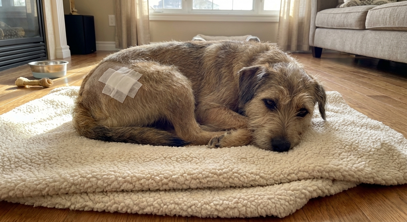
- Prevent licking or chewing at masses and incisions: Use an e-collar or recovery suit as needed.
- Keep a simple log: Appetite, energy, stools, and any changes in lumps.
If you want to explore fresh food or home-prepared options during cancer care, do it thoughtfully and with guidance. Cancer patients can have special needs, and the goal is always safe, balanced nutrition that your dog can tolerate.
Questions to ask your veterinarian or oncologist
- Was the tumor graded using Patnaik, Kiupel, or both?
- Were margins clean? If not, what are the next best options?
- Do we need lymph node aspirates or abdominal ultrasound?
- Is this location considered higher risk or harder to fully remove?
- Should we test for c-KIT mutation or review KIT staining?
- What signs of recurrence or spread should I watch for at home?
- What is the goal of treatment: cure, control, or comfort?
If you take only one thing from this page, let it be this: get new or changing lumps checked early. Many mast cell tumors can be managed very successfully when we act quickly and choose the right plan for the individual dog.
Sources
- American College of Veterinary Surgeons (ACVS). Client Education: Mast Cell Tumors. https://www.acvs.org
- American College of Veterinary Internal Medicine (ACVIM). Consensus Statements and oncology resources (mast cell tumor staging and treatment discussions). https://www.acvim.org
- Kiupel M, Webster JD, Bailey KL, et al. Proposal of a 2-tier histologic grading system for canine cutaneous mast cell tumors to more accurately predict biological behavior. Veterinary Pathology. 2011.
- Blackwood L, Murphy S, Buracco P, et al. European consensus document on mast cell tumours in dogs and cats. Veterinary and Comparative Oncology. 2012.
- Veterinary oncology reviews and textbooks covering prognostic factors such as mitotic index, KIT staining patterns, and c-KIT mutation status.