When someone tells me their cat has a “mast cell tumor,” the first thing I ask is a very calm follow-up question: Where is it? In cats, mast cell tumors (MCTs) often fall into two big buckets, skin and internal (most commonly the spleen or intestinal tract). The day-to-day experience for your cat, plus the treatment conversation you will have with your veterinarian, can look very different depending on which form you are dealing with.
As a veterinary assistant in Frisco, Texas, I have seen how scary the word “tumor” feels. My goal here is to help you separate what is typical for feline cutaneous (skin) mast cell tumors from visceral (internal) disease, and to walk you through what staging and treatment planning usually include.
What mast cells do
Mast cells are immune cells found throughout the body, especially in the skin, respiratory tract, and GI tract. They store chemicals like histamine and heparin that help with inflammation and allergic responses.
When mast cells turn cancerous and form a tumor, they can still release those chemicals. That is why mast cell tumors can cause symptoms that seem unrelated to a lump, such as:
- Itchiness or red, inflamed skin around a mass
- Swelling that waxes and wanes
- Vomiting, decreased appetite, or nausea (more common with internal disease)
- GI irritation and, in some cases, ulceration that histamine can contribute to (this is emphasized more consistently in dogs than cats, but it can occur, especially with visceral or systemic disease)
Not every cat shows these signs, but it helps explain why your vet may prescribe antihistamines or stomach support medications as part of the plan.
Skin MCTs
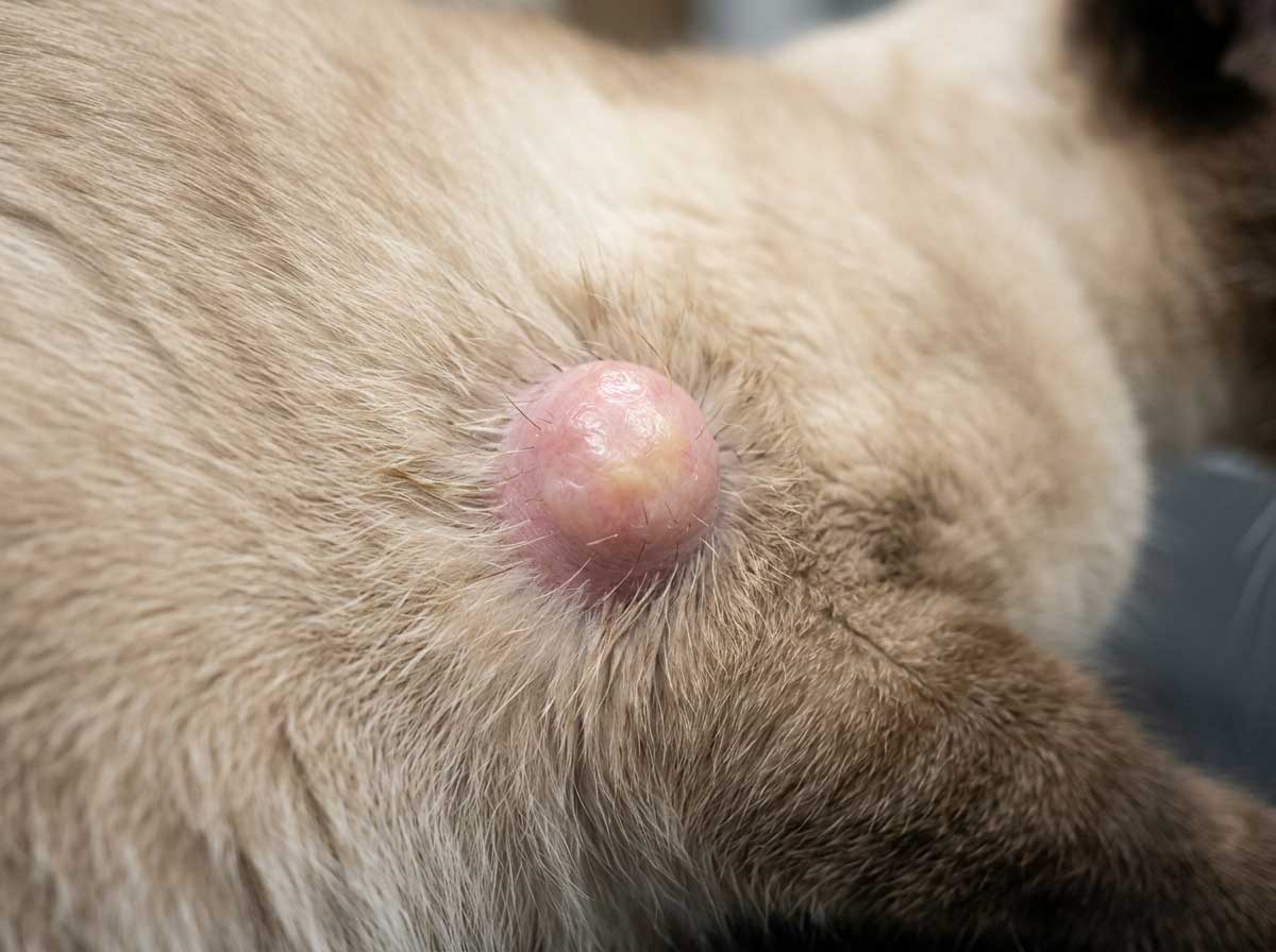
Cutaneous mast cell tumors are one of the more common round cell tumors of feline skin, although overall frequency can vary depending on the dataset and region. Many cats present with a small bump (or bumps) that an owner finds while petting.
Here is the big transition point to keep in mind: with skin MCTs, the story often starts with “I found a lump.” With visceral disease, it more often starts with “something is off.” That difference matters when you and your vet talk about testing and next steps.
Common looks
- A firm or rubbery nodule in or under the skin
- A hairless or slightly crusty patch
- A raised area that can look pink, tan, or pigmented
- Sometimes a lesion that seems to shrink and flare
In cats, skin MCTs can be solitary or multiple. They often show up on the head and neck, but they can occur anywhere.
One other nuance your vet may mention: feline skin mast cell tumors can come in different patterns under the microscope (often described as mastocytic versus histiocytic). In some younger cats, the histiocytic pattern is often more benign and can even regress. Mastocytic tumors can be more variable. Either way, it is not something to guess at without your veterinarian and a pathology report guiding the plan.
When people hear “spread,” they often picture lung metastasis right away. With many feline skin MCT cases, the more common worries are local recurrence at the surgical site or new skin lesions popping up over time, not immediate widespread metastasis. Your vet will still look for signs of broader disease, but the word “spread” does not always mean the worst-case scenario.
Internal mast cell disease
Internal (visceral) mast cell disease tends to change the whole feel of the case. Instead of “my cat has a bump,” owners often say “my cat is just not acting right.” Two major internal locations are the spleen and the intestines.
Splenic involvement
When mast cells involve the spleen, cats may develop:
- Weight loss
- Lethargy or hiding more
- Decreased appetite
- Vomiting (sometimes)
- A vet may feel an enlarged spleen on exam
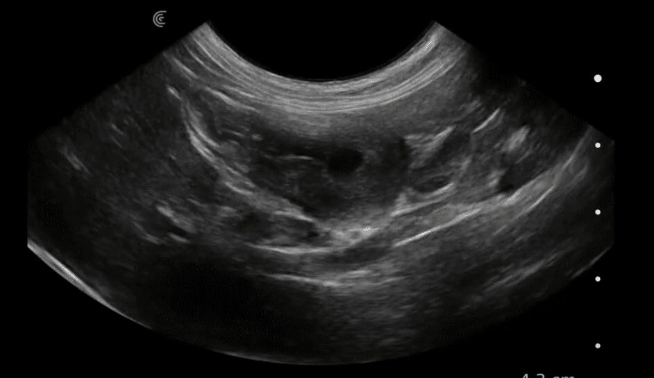
Many splenic cases are discovered after bloodwork or an ultrasound done for vague symptoms. Splenic disease can be the main problem, but it can also be part of a more systemic process, which is why your vet may recommend additional staging even after spleen surgery.
Intestinal MCT
Intestinal mast cell tumors can mimic other chronic GI diseases (including inflammatory bowel disease and lymphoma). Signs often include:
- Chronic vomiting or nausea
- Diarrhea or soft stool
- Weight loss despite eating
- Abdominal discomfort or a tucked-up posture
Because cats are so good at masking illness, subtle changes like “eating less,” “sleeping in a new spot,” or “not jumping up like usual” can be important clues.
One important detail here: a “mast cell” finding in the GI tract can be tricky. Cats can have mast cell-rich inflammation that is not cancer, and some cancers and inflammatory conditions overlap in how they look and behave. Definitive diagnosis often relies on histopathology (and sometimes special stains or immunohistochemistry), plus a pathologist interpreting the findings in context.
Cats vs dogs
If you have ever read about mast cell tumors in dogs, you may have seen detailed tumor grading systems used to predict behavior and guide treatment. That canine approach is not a clean fit for cats.
Here is the practical takeaway I share with families: feline mast cell tumors do not reliably follow the same grading rules as canine skin mast cell tumors. In cats, the conversation is usually less about a single “grade” number and more about:
- Location (skin vs spleen vs intestine)
- Whether the tumor appears solitary or part of broader mast cell disease
- Whether complete surgical removal is possible
- Whether there is evidence of involvement beyond the primary site
- Your cat’s overall health and comfort
That is also why you may hear your veterinarian focus on staging tests and imaging early in the process, especially if anything suggests internal involvement.
Diagnosis
Most cases start with a physical exam and a discussion of symptoms. From there, diagnostics depend on whether the concern is a skin mass or internal disease.
For a skin lump
- Fine needle aspirate (FNA): a tiny needle collects cells for a quick look under the microscope. Mast cells often exfoliate well, so FNAs can be very helpful.
- Biopsy and histopathology: if removal is planned, the mass is sent to a lab to confirm the diagnosis and evaluate margins.
A practical point: mast cell tumors can “react” when handled, meaning they can get temporarily swollen, red, or itchy due to mediator release. Big, dramatic reactions are talked about more in dogs than cats, but many clinicians still take reasonable precautions. Depending on the location, your cat’s history, and what is being done (sampling vs surgery), your vet may recommend pre-medications such as an H1 antihistamine and sometimes additional GI support. This is very case-by-case and not a universal requirement.
For suspected internal disease
- Bloodwork (CBC and chemistry) to look for anemia, organ changes, and overall health status
- Abdominal ultrasound to evaluate spleen, intestines, liver, and lymph nodes
- FNA of spleen or lymph nodes when appropriate
- GI biopsies (endoscopic or surgical) when intestinal disease is suspected and imaging is not enough
If you are anxious about timing, you are not alone. FNAs are sometimes read the same day (especially in-clinic), while biopsy results typically take days once the lab receives the sample. Your vet team can usually tell you what to expect for your specific clinic.
Staging
Staging means checking how far the disease goes and what organs are involved. For cats, staging is customized to the individual case, but the discussion often includes:
- Measuring and mapping skin masses and checking for additional nodules
- Palpating lymph nodes and sampling them if enlarged or suspicious
- Abdominal imaging if there are GI signs, weight loss, or concern for splenic involvement
- Chest imaging when your vet wants a broader health picture (anesthesia planning, ruling out other causes of symptoms, and case-by-case screening when visceral disease or systemic signs raise concern). For feline cutaneous MCT, lung metastasis is generally considered uncommon, so thoracic imaging is often more situational than automatic.
- Pathology review of removed tissue, including margin assessment if surgery is performed
If your cat has a skin mast cell tumor but is otherwise thriving, your vet may stage more lightly. If your cat has weight loss, vomiting, anemia, or an enlarged spleen, staging tends to be more thorough.
Treatment options
Treatment depends heavily on whether the tumor is in the skin or internal organs. The best plan is the one that treats the disease while also protecting your cat’s quality of life.
Skin MCTs
- Surgery: often the first choice for a solitary skin tumor that can be removed.
- Histopathology: helps confirm diagnosis and whether the surgeon got it all. In cats, margin guidance is not always as standardized as canine MCT rules, but margin information is still useful for planning what comes next.
- Monitoring: some cats develop more than one skin lesion over time, so rechecks matter.
Depending on the case, your vet may discuss additional therapies if a tumor cannot be fully removed, if there are multiple lesions, or if the pathology suggests more aggressive behavior.
Splenic involvement
- Splenectomy (spleen removal): commonly discussed when the spleen is the primary site and imaging supports surgical treatment. Many cats do surprisingly well after recovery.
- Next steps after surgery: the spleen is sent for histopathology, and your vet may recommend additional staging (for example, checking liver or bone marrow) if there are concerns about systemic disease.
- Medical management: antihistamines, GI support, and sometimes chemotherapy depending on staging and clinical signs.
Intestinal MCT
- Surgery: may be considered if there is a localized intestinal mass that can be removed.
- Chemotherapy: often part of the conversation for intestinal disease, especially if diffuse involvement is suspected.
- Supportive care: appetite support, anti-nausea medication, GI protectants, pain control, and nutrition planning are not “extras.” They are central.
For visceral disease, referral to oncology or internal medicine can be very helpful. A specialist may add advanced imaging, deeper biopsy planning, and a chemo and supportive-care roadmap tailored to your cat.
Prognosis
With feline mast cell tumors, prognosis is less about a one-size-fits-all number and more about a few key questions:
- Is it skin-only, or is it internal? Skin-only disease that is fully removed can have a much better outlook than intestinal disease.
- Was the tumor completely removed? Clean margins after surgery are a good sign for a solitary skin mass.
- Is the spleen the main issue? Some cats with splenic disease can do well after splenectomy, especially when no other sites are involved.
- How sick is the cat right now? Weight loss, poor appetite, anemia, and ongoing vomiting or diarrhea can signal a heavier disease burden.
- What did staging show? Lymph node involvement, liver changes, or widespread GI involvement can change the plan.
If your veterinarian recommends an oncology consult, it is not because they are giving up. It is because mast cell disease can be nuanced, and a specialist can help tailor treatment to your cat’s specific presentation and your goals for care.
At-home clues
Cats rarely read the textbook, and they often whisper instead of shout. If you are watching a cat with a known or suspected mast cell tumor, keep notes on:
- Appetite (including “walking away” after a few bites)
- Vomiting frequency and what it looks like
- Stool quality and litter box habits
- Energy, hiding, and social behavior
- Grooming (less grooming can be a sign they feel unwell)
- Weight (a baby scale can be very helpful)
For skin disease, also track the size, redness, and itchiness around the lump. If a mass suddenly swells, reddens, or bothers your cat, call your vet. Mast cell tumors can flare due to local inflammation and mediator release.
Questions to ask
- Does this look like cutaneous or visceral mast cell disease based on exam and initial tests?
- Do you recommend FNA, biopsy, or straight to surgical removal?
- What staging tests do you recommend and why (bloodwork, ultrasound, lymph node sampling, chest imaging)?
- If surgery is planned, what are the goals for margins and what would we do if margins are not clean?
- Should my cat be on antihistamines or GI support medications before or after procedures?
- What signs at home would be an emergency for you?
- Would an oncology referral change the plan for my cat?
When to seek urgent care
Please call your vet or an emergency clinic right away if your cat has any of the following:
- Repeated vomiting with inability to keep water down
- Black, tarry stool or obvious blood in stool
- Sudden collapse, extreme weakness, or pale gums
- Rapidly enlarging mass with significant swelling or pain
- Labored breathing
These signs do not automatically mean the tumor is worse, but they do mean your cat needs help quickly.
A hopeful note
I have met many cats with skin mast cell tumors who go right back to being themselves after surgery. I have also supported families through internal mast cell disease where the path is more complex. In both situations, the best next step is the same: get clear on which form you are dealing with, complete the staging your veterinarian recommends, and build a plan that protects your cat’s comfort while targeting the disease.
If you want, tell me what your vet has said so far and what signs you are seeing at home (skin lump location, appetite, vomiting, weight loss). I can help you organize the right questions for your next visit.