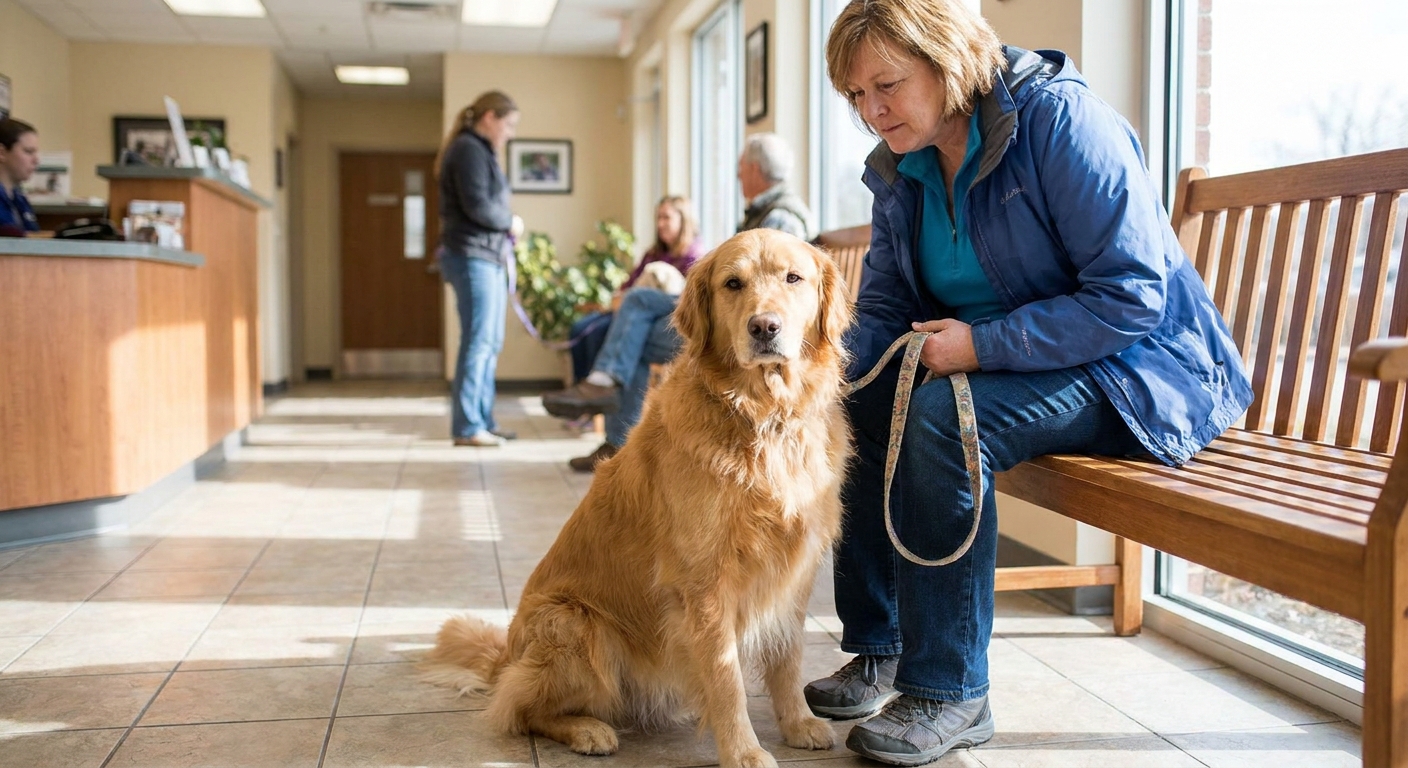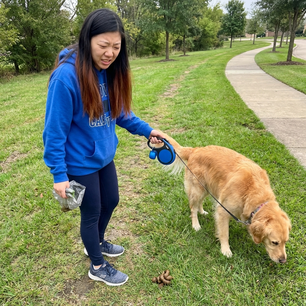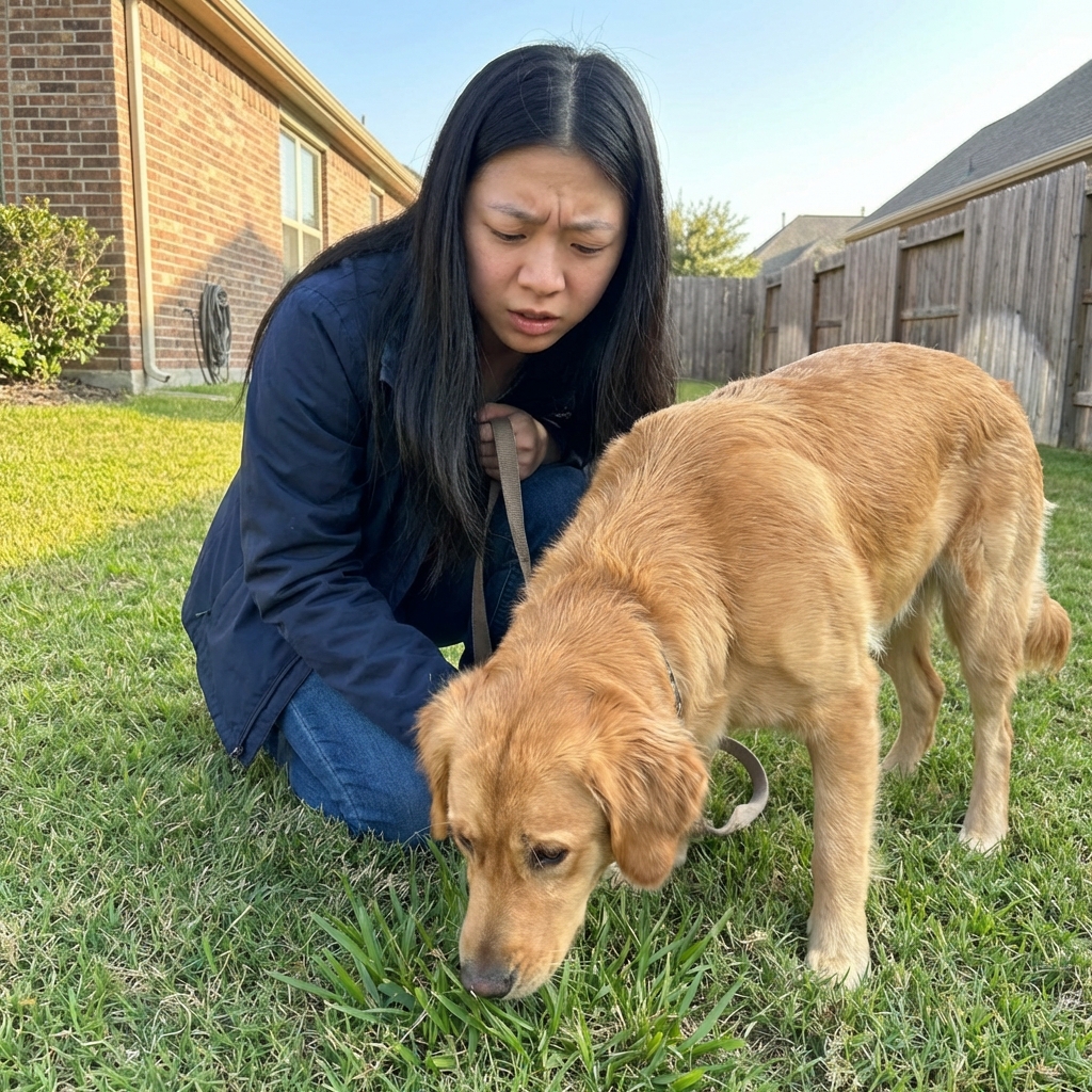
I have talked with a lot of worried pet parents who start with one scary symptom: “My dog is pooping mucus.” Sometimes that really is a one-off irritation that settles down in a day or two. But sometimes mucus is part of a much bigger intestinal problem, especially when it shows up alongside weight loss, ongoing diarrhea, or a suddenly swollen belly.
One condition that deserves a clear, separate explanation is intestinal lymphangiectasia. It is not the same thing as typical colitis, and it is not the same as general inflammatory bowel disease (IBD), even though signs can overlap. Lymphangiectasia is a protein-losing disease, and that detail changes how veterinarians diagnose it and how we talk about diet.
What it is
Lymphangiectasia means the lymphatic vessels are dilated. In the intestines, these tiny lymph vessels are called lacteals. Their job is to transport fluid, immune cells, and especially dietary fats away from the gut.
When lacteals become enlarged and leaky, the intestine can start losing:
- Protein (especially albumin) into the GI tract
- Fluid
- Lymphocytes (important immune cells)
- Fat-soluble vitamins (A, D, E, K) and other nutrients, depending on severity
This is why lymphangiectasia is often discussed under the umbrella term protein-losing enteropathy (PLE). Not every PLE is lymphangiectasia, but lymphangiectasia is a classic cause.
Primary vs secondary causes
Some dogs appear to have a more “primary” lymphatic problem (meaning the lymph vessels themselves are the main issue). In other dogs, lymphangiectasia is secondary, meaning something else is driving lymphatic congestion, inflammation, or obstruction.
Depending on the dog, veterinarians may look for underlying contributors such as:
- Chronic intestinal inflammation (including IBD)
- Intestinal lymphoma or other infiltrative disease
- Right-sided heart disease or conditions that raise venous pressure
- Liver disease or portal hypertension
- Severe intestinal infections or other less common inflammatory triggers
I am mentioning this because it answers the question almost everyone asks: “Why did this happen?” Sometimes we find a clear driver, and sometimes we do not. Either way, knowing what is fueling the protein loss guides treatment.
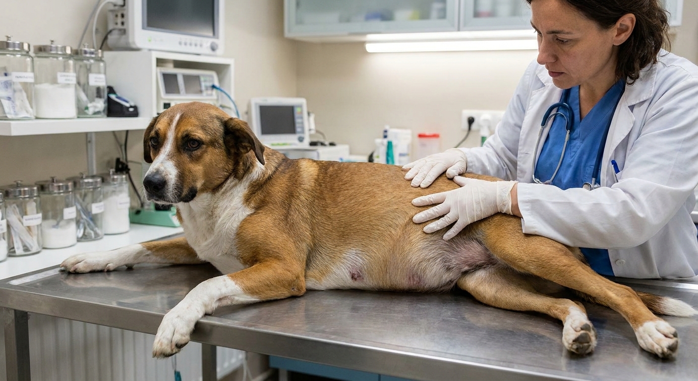
Why the belly can swell
When blood protein, particularly albumin, drops too low, the body cannot hold fluid inside blood vessels as effectively. Fluid can seep into tissues and body cavities, leading to:
- Ascites (fluid in the abdomen), which can look like a pot-bellied or suddenly swollen belly
- Peripheral edema (puffiness in legs or under the skin) in some dogs
- Pleural effusion (fluid around the lungs) less commonly, but it can happen
At the same time, the intestinal disease itself can cause chronic diarrhea, poor appetite, nausea, and weight loss.
Common symptoms
Signs can range from subtle to very serious. Many dogs do not show every symptom at once.
Digestive signs
- Chronic diarrhea, often soft or watery
- Greasy-looking stool in some cases (fat malabsorption)
- Mucus in stool can occur, but it is not the hallmark sign
- Vomiting in some dogs
- Reduced appetite or picky eating
Whole-body signs tied to protein loss
- Weight loss or muscle loss
- Lethargy, low energy
- Swollen belly from fluid accumulation
- Swelling in limbs or under the skin in some dogs
Something many families miss at first
Some dogs with lymphangiectasia do not look “thin” in the beginning because their belly is enlarged from fluid. You can have a dog losing muscle while also looking round in the abdomen.
Breeds at higher risk
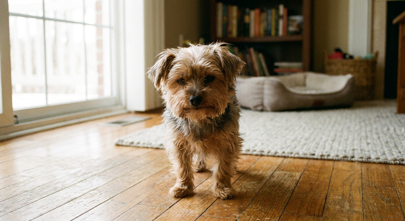
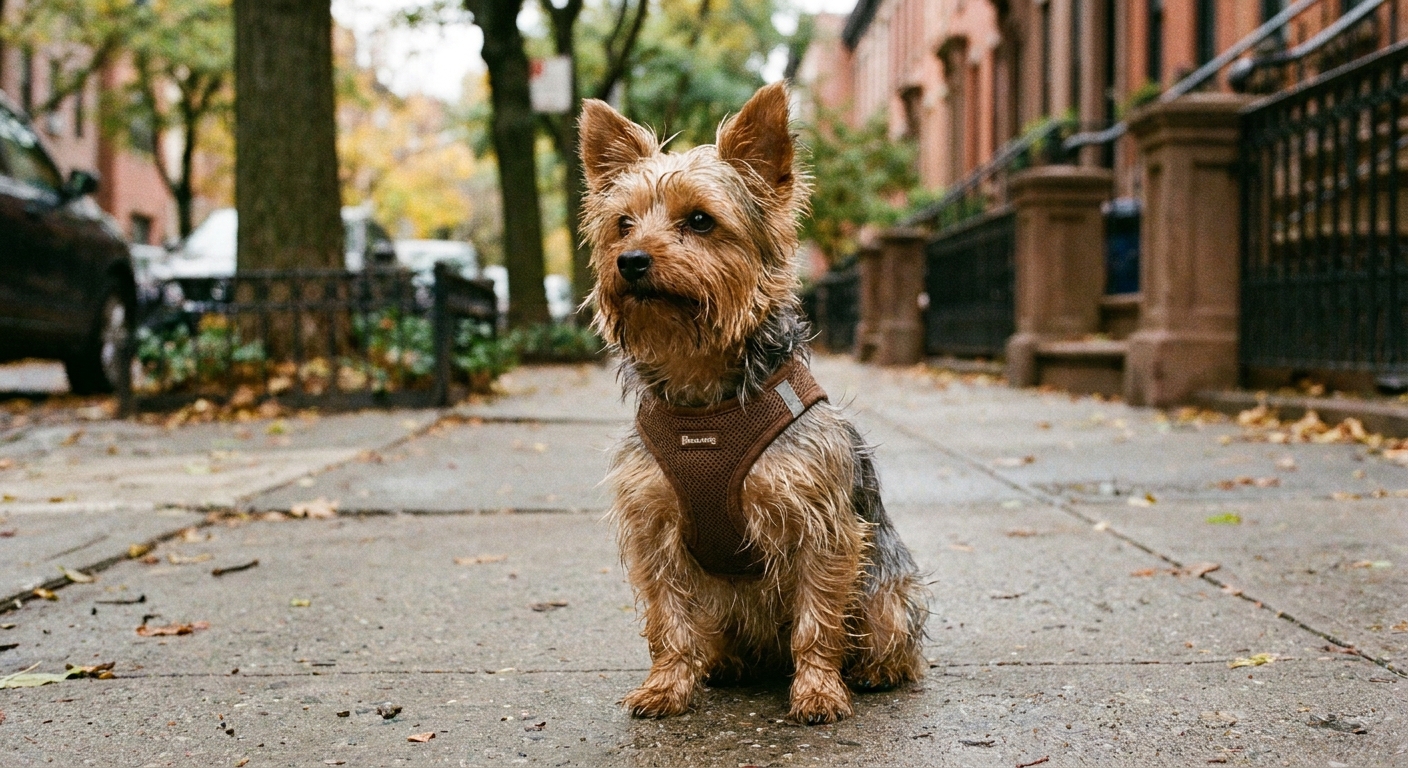
Lymphangiectasia can happen in any dog, but we do see patterns. It is often diagnosed in small breeds, including Yorkshire Terriers in particular. It can also be seen in other breeds (and mixed breeds), and it may appear alongside other intestinal conditions.
Mucus stool vs lymphangiectasia
I want to say this gently because I know how stressful poop changes can be.
One mucus stool after something like diet indiscretion, a stressful event, a minor GI bug, or a new treat is common. Mucus often goes along with large-bowel irritation (colitis), while lymphangiectasia is more often a small-intestinal problem. That said, the gut does not always read the textbook, and overlap happens.
If your dog is otherwise bright, eating, and acting normal, your vet may recommend monitoring, a bland diet for a short period, and watching hydration.
Lymphangiectasia is different. It tends to be a pattern, not a single episode. Red flags that suggest you should call your veterinarian promptly include:
- Diarrhea that is recurring, worsening, or not improving over 24 to 48 hours, especially if your dog seems unwell
- Repeated mucus stools over several days
- Weight loss
- Decreased appetite, vomiting, or lethargy
- Swollen belly or sudden abdominal enlargement
- Weakness, collapse, pale gums, or trouble breathing
How vets test for it
Diagnosis is usually a combination of history, lab work, imaging, and sometimes endoscopy and biopsies. The goal is to confirm the protein-losing problem and identify the underlying cause.
1) Bloodwork
- Total protein and albumin: often low
- Globulins: may be low too
- Cholesterol: sometimes low (it can be normal in some cases)
- Lymphocyte count: can be low (lymphopenia)
- Electrolytes, kidney and liver values: to assess overall status and rule out other causes
Depending on the case, your veterinarian may also discuss calcium. When albumin is low, total calcium can look low too, even when the biologically active portion is normal. Your vet can interpret this in context.
2) Urinalysis (to rule out kidney protein loss)
Protein can also be lost through the kidneys (protein-losing nephropathy). A urinalysis and sometimes a urine protein-to-creatinine ratio help separate intestinal loss from kidney loss.
3) Fecal testing
Vets often check for intestinal parasites and infections because chronic diarrhea has many causes. This is especially important if a dog is not on reliable parasite prevention.
4) Imaging
- Abdominal ultrasound can show intestinal changes and fluid in the belly. In some cases, ultrasound findings can strongly raise suspicion for lymphangiectasia.
- X-rays may be used to assess abdominal distension or rule out other problems.
5) GI panels and specialized tests
Depending on the case, your veterinarian may recommend tests such as:
- cobalamin (B12) and folate levels
- pancreatic testing to rule out exocrine pancreatic insufficiency (EPI) or pancreatitis concerns
- fecal alpha-1 proteinase inhibitor testing in some settings, which can help confirm protein loss through the intestines
6) Endoscopy and biopsy
When a definitive answer is needed, the confirmatory standard is usually intestinal biopsy (endoscopic or surgical) with histopathology. Biopsy can also help determine whether inflammatory disease, lymphoma, or other intestinal conditions are present at the same time.
One nuance that is worth knowing: lymphangiectasia can involve deeper layers of the intestine, so in some dogs, endoscopic biopsies may not capture the full picture. Your veterinarian or internal medicine specialist can advise whether endoscopic samples are appropriate or whether full-thickness biopsies are more useful in your dog’s situation.
If your dog has a very low albumin, your veterinarian may recommend stabilizing first before more invasive testing. Safety always comes first.
The very-low-fat diet
This is the heart of lymphangiectasia management.
Because the intestinal lymphatics transport fats, a high-fat meal can increase lymph flow and pressure inside those dilated lacteals. In dogs with lymphangiectasia, that can mean more leaking and worsening protein loss.
That is why veterinarians commonly recommend a very-low-fat diet as a cornerstone of treatment. Not “low fat compared to average.” Very low.
What “very low fat” means
Targets vary by veterinarian and by the dog’s lab values, but the overall goal is strict fat control, typically using diet labels your vet can compare on an equal basis (often grams of fat per 100 kcal).
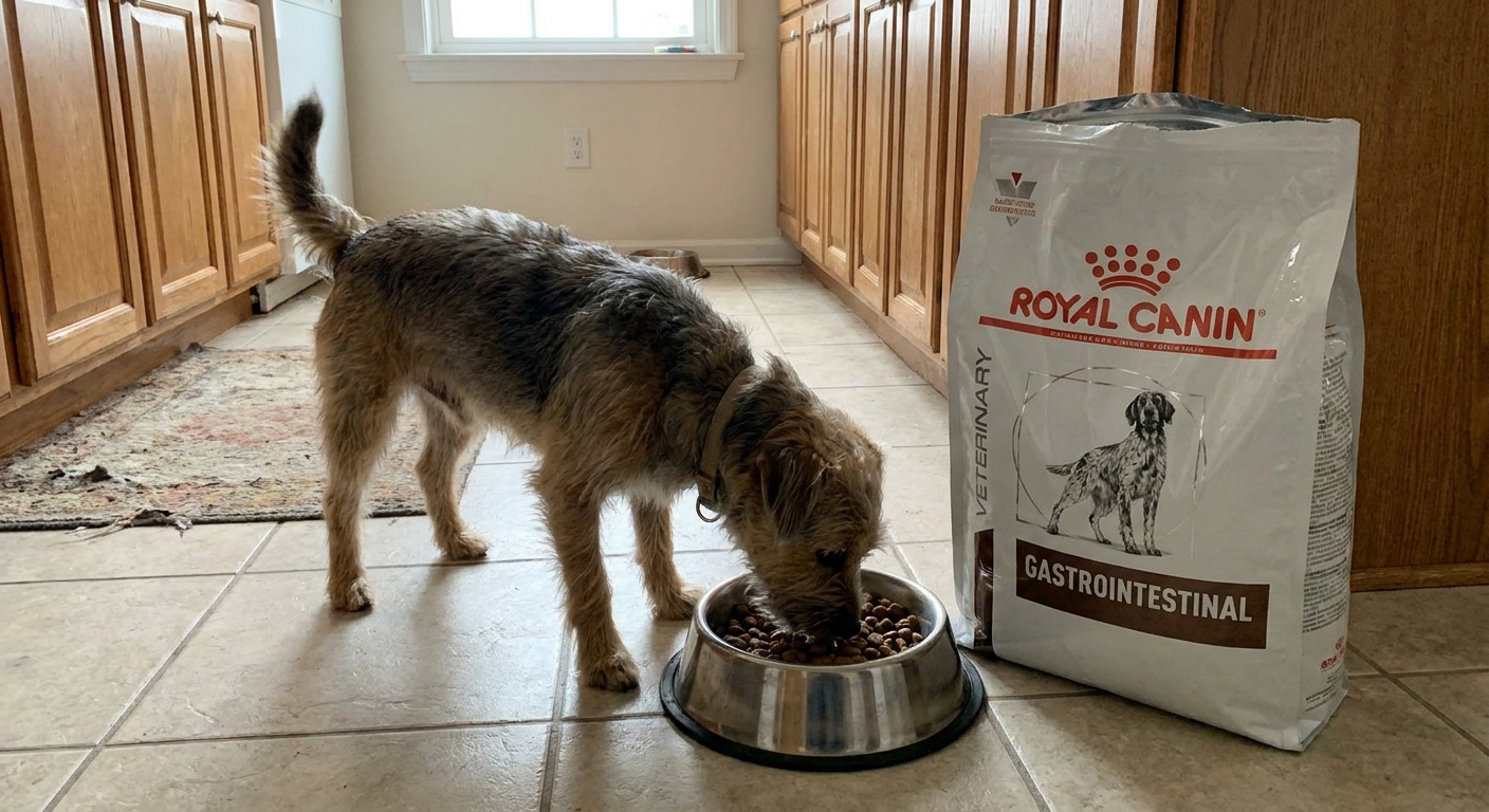
This often requires a prescription gastrointestinal diet, at least initially, because these diets are formulated to hit specific nutrient targets while staying extremely low in fat.
Important caution about homemade diets
As someone who loves whole foods and has seen how powerfully nutrition can support pets, I still want to be clear: lymphangiectasia is not a DIY diagnosis or a DIY diet situation.
Homemade diets for PLE can be done, but they should be built with your veterinarian and ideally a board-certified veterinary nutritionist. The margin for error is small, especially when your dog is already low in albumin, losing nutrients, or has other conditions happening at the same time.
Other treatments
Diet is often step one, but not always the only step.
- Medications to reduce intestinal inflammation if inflammatory disease is present (your vet may discuss steroids or other immunosuppressive medications depending on the diagnosis)
- Vitamin and mineral support such as cobalamin (B12) supplementation when low
- Probiotics in some cases, based on the dog’s GI history
- Careful fluid management if your dog is dehydrated from diarrhea or has significant fluid shifts
- Plasma or albumin products, or other oncotic support in select severe cases, when your veterinarian feels the potential benefits outweigh the risks
- Blood clot prevention in some higher-risk patients, because protein-losing states can increase clot risk (this is individualized)
The exact plan depends on what is driving the lymphangiectasia and how sick your dog is right now.
Other causes of PLE
Another important point: lymphangiectasia is not the only reason dogs develop protein-losing enteropathy. Your veterinarian may also consider possibilities such as:
- Severe inflammatory bowel disease
- Intestinal lymphoma or other intestinal cancers
- Severe intestinal ulceration or erosive disease
- Some systemic or infectious diseases that affect the gut
This is one reason the diagnostic plan can feel broad at first. Your vet is trying to confirm protein loss, then narrow down the “why” so treatment is targeted.
Monitoring and follow-up
Lymphangiectasia is a monitor-and-adjust condition. Even when a dog improves clinically, follow-up helps make sure the protein loss is truly under control.
Monitoring often includes:
- Rechecking albumin and total protein (and sometimes cholesterol and lymphocyte counts)
- Tracking weight, muscle condition, and stool quality
- Adjusting diet and medications based on response
- Rechecking ultrasound or belly fluid status in some dogs with ascites
Prognosis
The outlook varies a lot.
Some dogs do very well long-term with consistent diet management and periodic monitoring. Others have more stubborn disease, especially if lymphangiectasia is secondary to another serious intestinal condition.
The best thing you can do is stay partnered with your veterinarian and keep follow-up appointments. Lymphangiectasia is one of those conditions where we learn a lot from repeat lab work and from how your dog responds to diet changes.
When to seek urgent care
Please contact your veterinarian urgently or go to an emergency clinic if you notice:
- Severe lethargy or collapse
- Difficulty breathing (fluid around lungs can be life-threatening)
- A rapidly enlarging abdomen
- Profuse watery diarrhea, black stool, or bloody diarrhea
- Repeated vomiting with inability to keep water down
- Signs of dehydration (tacky gums, sunken eyes, weakness)
FAQ
Can lymphangiectasia cause mucus in stool?
Yes, it can. But mucus by itself is not specific to lymphangiectasia. The bigger clue is the overall picture, especially chronic diarrhea, weight loss, and lab work showing low protein or low albumin.
Is lymphangiectasia the same as IBD?
No. IBD is a broad term for chronic intestinal inflammation. Lymphangiectasia is a problem with the intestinal lymphatics becoming dilated and leaky. A dog can have both, and biopsies can help sort out what is happening.
Why does my dog look bloated if they are losing weight?
A distended belly can be due to fluid (ascites) from low albumin. Meanwhile, the dog may be losing muscle and body condition underneath. This is one reason an exam and lab work are so important.
Will changing food help right away?
Some dogs improve quickly with the right very-low-fat diet, but others take longer and need additional treatment. Your vet will usually recheck blood protein values to confirm the plan is working.
A supportive next step
If you are reading this because your dog has ongoing diarrhea, mucus stools that are not resolving, or a belly that looks swollen, please do not blame yourself. GI conditions can be sneaky, and lymphangiectasia is not something most families would recognize on their own.
Call your veterinarian and ask directly whether protein-losing enteropathy is on the list, and whether checking albumin and total protein makes sense for your dog. Getting the right diagnosis early can make the diet conversation clearer and, most importantly, can help your pup feel better sooner.