

Discover a new lump on your dog’s back? Follow calm, vet-smart steps: check size and skin changes, rule out ticks, prevent licking, know urgent signs, and ...
Article
•
Designer Mixes
Lumps on Dogs: Lipomas vs. Mast Cell Tumors

Shari Shidate
Designer Mixes contributor
Finding a new lump on your dog can turn your stomach in a second. I get it. As a veterinary assistant, I have watched countless families go from “It’s probably nothing” to “Please tell me what it is” in one appointment.
The good news is that many lumps are benign, and one of the most common is a lipoma (a fatty growth). The not-so-simple truth is that some lumps can be mast cell tumors, and those can look like almost anything. This article will help you understand what to watch for, what you can do at home, and when to get your veterinarian involved.

First things first: You cannot diagnose a lump by touch alone
There are clues that suggest a lipoma versus a mast cell tumor, but there is no reliable way to confirm the difference at home. Even experienced veterinary teams do not guess. We test.
Also, it is not always a lipoma or a mast cell tumor. Dogs can get other common lumps too, like sebaceous or follicular cysts, warts (papillomas), abscesses, hematomas, enlarged lymph nodes, or other skin tumors. The outside does not reliably tell you the inside.
In most general-practice clinics, the most useful first test for many skin lumps is a fine needle aspirate (FNA). Your veterinarian uses a small needle to collect cells from the lump, then examines them under a microscope in-house or sends slides to a lab. It is quick, usually low-stress, and often less expensive than surgery.
If your dog has a new lump, a changing lump, or a lump that worries you, ask your vet about an FNA. Knowing is almost always less stressful than wondering.
What is a lipoma?
A lipoma is a benign tumor made of fat cells. They are extremely common, especially in middle-aged and senior dogs. Many dogs live their whole lives with one or more lipomas that never cause problems.
Common lipoma clues
- Soft and squishy, like a little pouch of dough
- Moves easily under the skin when you gently push around it
- Usually not painful
- Slow-growing over months to years
- Often on the chest, belly, armpits, or upper legs
Two important caveats: some lipomas can feel firmer (especially if they are more fibrous), and some malignant tumors can also feel soft or movable. That is why we do not rely on feel alone.
Some lipomas can grow large and get in the way of movement, especially if they sit near an armpit or groin. There is also a type called an infiltrative lipoma that can grow into surrounding tissues. These are still made of fat cells, but they can be harder to remove completely. Imaging and biopsy are sometimes part of the workup, and recurrence after surgery can happen.
What is a mast cell tumor?
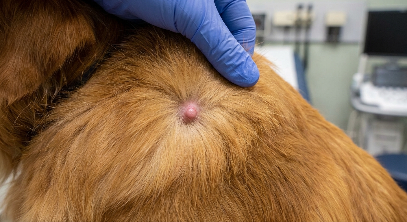
Mast cell tumors (MCTs) are among the most common malignant skin tumors in dogs. Mast cells are part of the immune system. When they become cancerous, they can form tumors in the skin or under the skin and may release histamine and other inflammatory chemicals.
Here is the tricky part: mast cell tumors are called “the great pretenders” because they can mimic many other types of lumps, including benign ones.
Common mast cell tumor clues
- Changes in size, sometimes swelling up and shrinking (even within days)
- Redness, irritation, or itching around the lump
- May feel firm or may feel soft, depending on the tumor
- May ulcerate (open sore) or look like a bug bite that will not heal
- May be solitary or multiple
Because mast cell tumors can release histamine, some dogs also show signs like vomiting, diarrhea, poor appetite, or stomach ulcer irritation. Not every dog does, but it is something your vet may consider. This is also why veterinarians sometimes use antihistamines and, in some cases, stomach protectants alongside other treatment.
Lipoma vs. mast cell tumor
Use this as a general guide only. It is not a diagnosis.
- Feel: Lipomas are often soft and very movable. Mast cell tumors can be firm or soft and may feel “stuck” or may move.
- Speed: Lipomas tend to grow slowly. Mast cell tumors may change quickly or fluctuate in size.
- Skin changes: Lipomas usually do not change the skin. Mast cell tumors may cause redness, swelling, irritation, or ulceration.
- Behavior: Lipomas are usually quiet and boring. Mast cell tumors can be unpredictable, which is why diagnosis, grade, and margins matter.
Red flags: book the vet visit
I encourage families to trust their gut. If you are worried, that alone is a reason to schedule an exam. That said, these are the big medical “do not wait” signs:
- The lump is growing quickly or changing shape
- The lump shrinks and swells repeatedly
- The skin over it is red, warm, bleeding, or ulcerated
- Your dog is licking, scratching, or seems painful when touched
- The lump is near the mouth, genitals, armpit, or groin (areas where tumors can be harder to remove cleanly)
- Your dog seems unwell: vomiting, diarrhea, low appetite, lethargy
- Your dog has multiple new lumps appearing over a short period
When to seek urgent care
It is uncommon, but some reactions can be more serious. Seek urgent veterinary care if your dog has sudden facial swelling or widespread hives, repeated vomiting, weakness or collapse, trouble breathing, or black, tarry stool.
What to do at home
1) Start a simple lump log
This is one of the most helpful things you can do before your appointment.
- Write down the date you found it
- Note the location (example: “right side chest, 2 inches behind front leg”)
- Measure it with a ruler (in millimeters or inches) and record the size
- Note if it feels soft, firm, or fixed
- Track any changes weekly
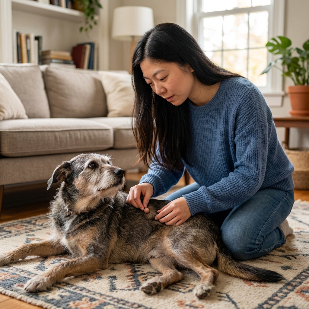
2) Take clear photos
Use good lighting and part the fur. Photos help you notice subtle changes and can help your vet if the lump looks different on appointment day.
3) Do not squeeze, lance, or pop it
Please do not try to drain a lump at home. If it is a mast cell tumor, irritation and pressure can trigger more inflammation, swelling, and bleeding. Even if it is not, poking at it can delay the right diagnosis and make the area harder to evaluate.
How vets confirm what a lump is
Fine needle aspirate (FNA)
As mentioned above, an FNA is often the best first step. It can identify fat cells (supporting lipoma) and it can sometimes clearly identify mast cells (raising concern for MCT).
One important limitation: FNAs do not always give a clear answer. Sometimes the sample does not collect enough diagnostic cells, and sometimes a lump has mixed tissue so the needle misses the most informative spot. If results are unclear or the lump is still suspicious, the next step is usually biopsy.
Biopsy and histopathology
Histopathology (examining tissue under a microscope) is the diagnostic gold standard. Your veterinarian may recommend a biopsy or surgical removal with submission to a pathologist. For mast cell tumors, the pathology report can include tumor grade (often reported as high vs low grade) and other features that guide next steps and prognosis.
Staging tests (when needed)
If a mast cell tumor is confirmed, your vet may discuss tests such as lymph node sampling, bloodwork, abdominal ultrasound, or additional imaging depending on the case.
Treatment overview
Lipomas
- Monitor if small and not interfering with movement
- Surgical removal if rapidly enlarging, limiting mobility, or bothersome
- Weight management can help overall health, but it does not “dissolve” a lipoma
Mast cell tumors
- Surgery is often first-line when possible, with appropriate margins
- Additional treatment may include medications, chemotherapy, or radiation depending on tumor behavior and location
- Supportive care may include antihistamines or stomach protectants, because mast cells can release histamine
Your dog’s plan should be individualized. Two dogs can both have “mast cell tumors” and still have very different treatment paths and outcomes.
Prevention and wellness notes
You cannot prevent every lump. But you can stack the odds in your dog’s favor by staying consistent with:
- Hands-on monthly checks at home (like a mini “nose to tail” exam)
- Yearly or twice-yearly wellness visits, especially for seniors
- Healthy body weight and muscle maintenance
- Evidence-based parasite prevention so bites and skin inflammation are less likely to muddy the picture
And if you are working on diet upgrades, go slowly and choose balanced options. Great nutrition supports the immune system and skin health, but it should always complement, not replace, proper diagnostics for lumps.
Bottom line
Many dog lumps are harmless, and lipomas are common. But mast cell tumors are common too, and they can look deceptively “normal.” When a lump is new or changing, the kindest thing you can do for your dog and your own peace of mind is to get it checked and ask about an FNA, with the understanding that some lumps still need biopsy for a final answer.
Before your appointment, bring the helpful details: your dog’s age, breed mix, where the lump is located, when you first noticed it, and any size changes or photos. That information makes it easier for your veterinary team to move quickly and confidently.
Medical note: This article is educational and not a substitute for veterinary diagnosis or treatment.