Found a new lump on your dog? Compare lipoma vs mast cell tumor clues, know the warning signs, and learn why an FNA or biopsy is the only way to confirm what...
Article
•
Designer Mixes
Lump on Your Dog’s Back

Shari Shidate
Designer Mixes contributor
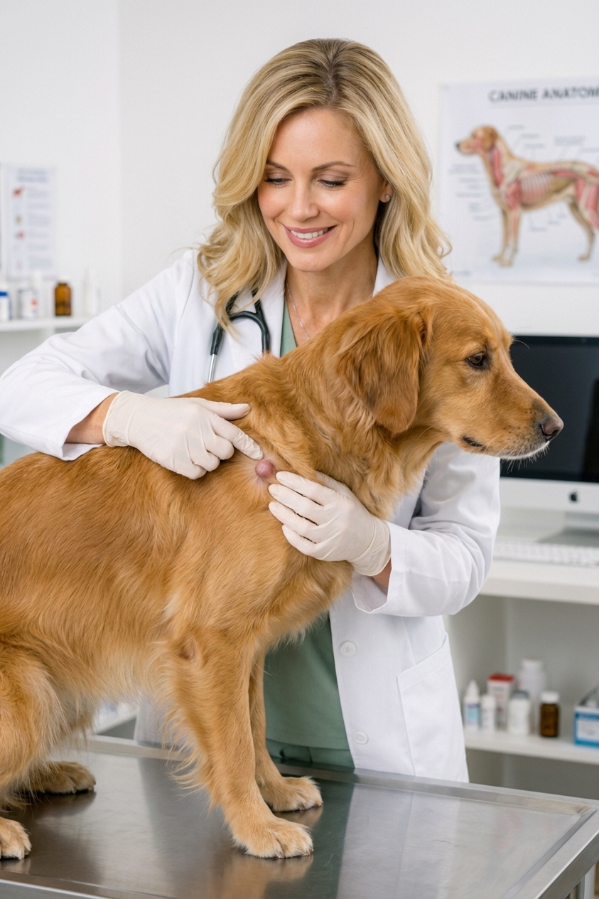
Finding a new lump on your dog’s back can feel scary. Take a breath. Many lumps turn out to be benign (not cancer), but some can be serious, and you cannot tell which by touch alone. Any new bump deserves a smart, calm plan. From my experience working in a veterinary clinic in Frisco, Texas, I have seen how much easier this is for families when they know what to watch, what to avoid, and when to book that vet visit.
This guide will help you make safe, evidence-based next steps while you wait for professional advice.

First steps at home
1) Do a quick, gentle check
- Location: Note exactly where it is (for example: mid-back, near the shoulder blades, right side).
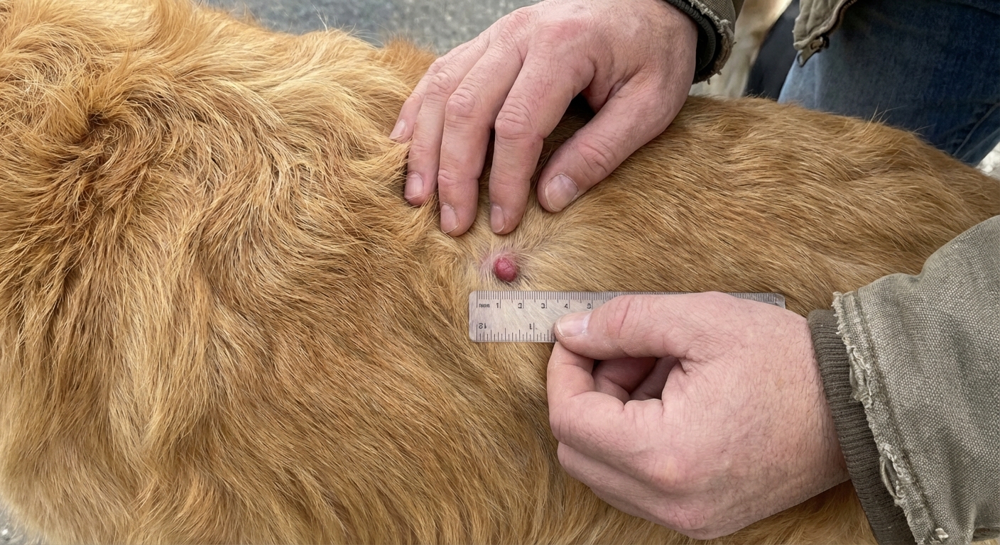
- Size: Measure it with a ruler or use a coin for scale. Write it down.
- Surface: Is the skin normal, red, scabby, moist, or bruised?
- Temperature and pain: Does it feel warm? Does your dog flinch?
- Mobility: With very light pressure, does it move under the skin or feel fixed in place?
- Number: Check for others. Multiple lumps can still be benign, but it changes your vet’s approach.
2) Take a clear photo today
Photos are incredibly helpful for tracking change. Take one close-up and one from a little farther away so the area is easy to identify later.
3) Make sure it is not a tick
Sometimes a “lump” is actually an attached tick. Part the fur and look closely. A tick is usually a distinct, round or oval body attached to the skin surface. If you are unsure, do not pull at it repeatedly. Call your vet for guidance on safe removal.
4) Prevent licking, chewing, and scratching
If your dog is bothering the area, an e-collar or soft cone can prevent irritation and infection. Avoid tight bandages on the back unless your vet instructs it.
5) Call your veterinarian and schedule an exam
If a lump is new, growing, firm, irregular, or your dog is acting “off,” it is worth scheduling an appointment soon. Many clinics start with an exam and often recommend a simple test called a fine needle aspirate (FNA) to help identify what the lump is made of.
What it might be
You cannot diagnose a lump at home, but knowing the usual suspects can reduce panic and guide good questions.
Benign possibilities
- Lipoma (fatty tumor): Often soft, movable, and slow-growing. Common in adult and senior dogs.
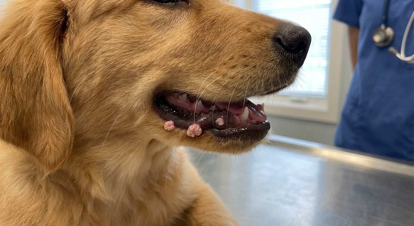
- Skin tag or wart (papilloma): Small growths from the skin surface. Can get irritated by collars, harnesses, or scratching.
- Sebaceous (follicular) cyst: A blocked hair follicle or gland that forms a lump. Sometimes it ruptures and leaks thick material.
- Swelling from trauma (contusion): A bump after rough play or a fall, sometimes tender and warm. Trauma-related swelling can mimic a true mass, especially early on.
- Abscess: A pocket of infection, sometimes from a bite or puncture. Often painful and warm, and it can worsen quickly. Some abscesses rupture and drain, and a fever may or may not be present.
More serious possibilities
- Mast cell tumor: Can look like almost anything. Some change size from day to day or look more swollen after being touched or scratched. This is one reason vets recommend testing instead of guessing.
- Soft tissue sarcoma: Often firm and may feel more attached under the skin.
- Other tumors: There are many skin and subcutaneous tumors in dogs. The good news is that early evaluation gives you the most options.
If you remember only one thing: new lump plus time equals uncertainty. A quick veterinary check and a needle sample are often the fastest way to get real answers.
When it is urgent
Go to an emergency clinic or urgent care the same day if you notice any of the following:
- Fast growth over hours to days
- Bleeding, oozing, or an open sore
- Sudden swelling of the face or hives (possible allergic reaction)
- Severe pain or your dog will not let you touch the area
- Fever, lethargy, not eating, vomiting, or your dog “just seems wrong”
- Difficulty breathing or collapse
What your vet may do
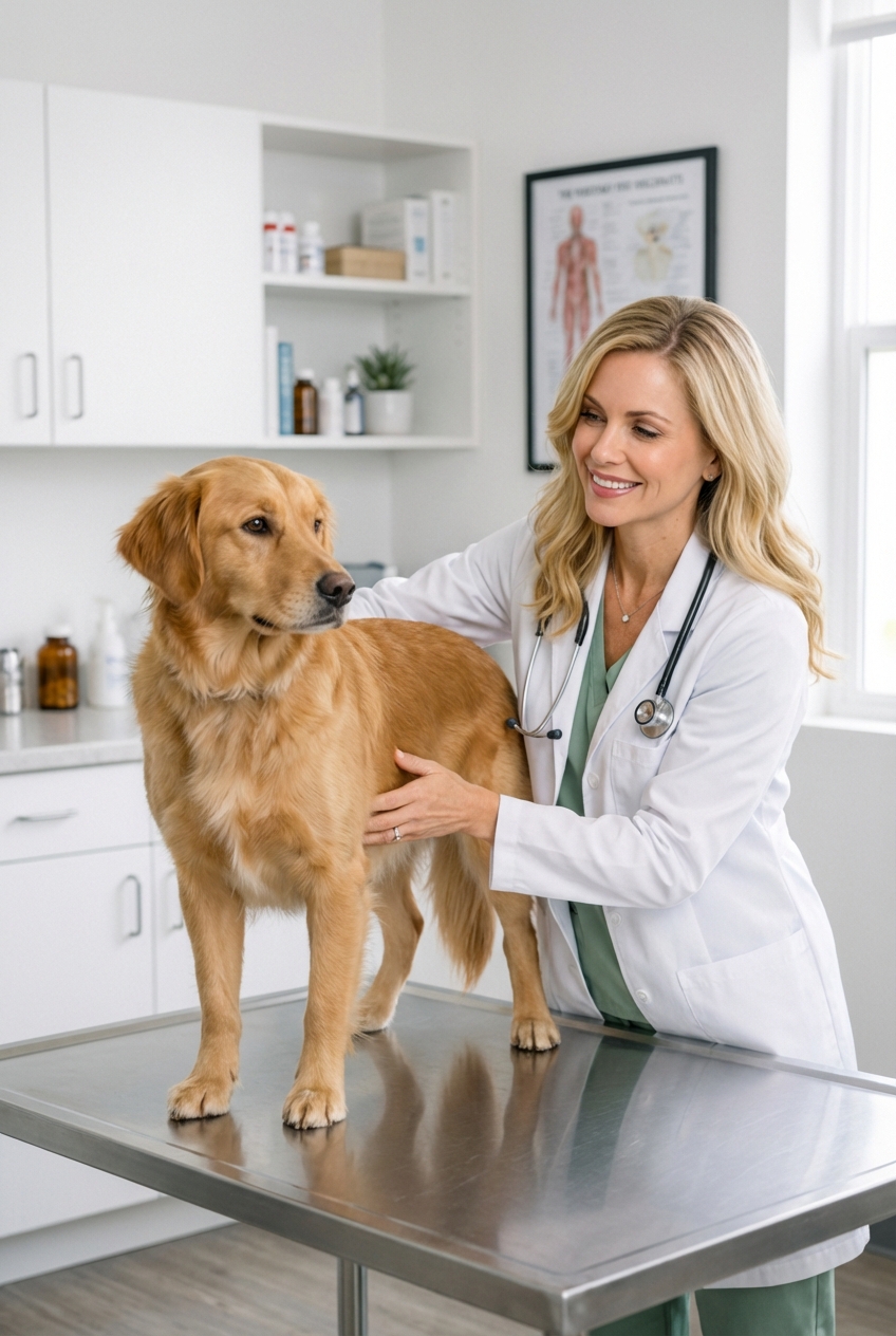
Physical exam
Your veterinarian will assess size, location, and whether it involves the skin, the tissue under the skin, or deeper structures.
Fine needle aspirate (FNA)
An FNA uses a small needle to collect cells. It is quick, minimally invasive, and often does not require sedation. That said, some dogs may need mild sedation if they are very anxious, if the area is painful, or if the lump is in a tricky location. Also, some lumps do not exfoliate cells well, so an FNA can be non-diagnostic even when everyone does everything right.
Biopsy or removal
If the FNA is inconclusive, or if the lump has concerning features, your vet may recommend a biopsy or surgical removal. The removed tissue is typically sent to a lab for histopathology, which is the most definitive way to identify the mass.
Imaging
In some cases, ultrasound helps show how a soft tissue mass extends and whether it involves deeper layers. X-rays are more useful when there is concern about bone involvement or when your vet wants to check the chest as part of screening or staging for certain cancers.
Avoid at-home fixes
I totally understand the urge to help right away, but these choices can make things worse or delay the right diagnosis.
- Do not squeeze, lance, or pop a lump. Cysts and abscesses can look similar from the outside, and squeezing can spread infection or inflammation.
- Do not apply essential oils or harsh topical products. Many are irritating or toxic to pets if licked.
- Do not give human pain medications like ibuprofen, naproxen, or acetaminophen unless your veterinarian has specifically prescribed them for your dog.
- Do not “watch it” indefinitely if it is changing. A short tracking window can be reasonable for some bumps, but growth, redness, pain, or ulceration should move the timeline up.
How to monitor well
Use a simple log
- Date found
- Size (in millimeters or centimeters)
- Texture (soft, firm, rubbery)
- Mobility (moves freely, slightly mobile, fixed)
- Skin changes (normal, red, crusty, hair loss)
- Your dog’s behavior (normal appetite and energy, or not)
A simple 1-2-3 guideline
Veterinary oncology groups often share a “see something, do something” style guideline. A commonly used 1-2-3 rule of thumb is to schedule an exam if a lump is:
- 1 month and still present
- 2 cm (about the size of a grape) or larger
- 3 months and growing
Also, do not wait on the timeline if it is rapidly changing, painful, bleeding, or the skin looks infected. When in doubt, earlier is fine.
Care while you wait
While you are waiting to be seen, focus on comfort and prevention.
- Keep the area clean and dry. If the skin is irritated, ask your clinic before applying anything.
- Prevent rubbing or trauma. Swap a harness that rubs the area for a different style, if needed.
- Support overall health. A balanced diet and a healthy weight matter. Vet-approved omega-3s may help support skin and coat health and may help some inflammatory conditions. If your dog is on prescription food or has a pancreatitis history, ask before adding supplements.
- Keep activity normal unless your dog seems sore. If it looks like a bruise or swelling from trauma, avoid rough play until assessed.
What to bring
- Your photos (including a size reference if possible)
- Your notes and timeline (when you first noticed it and whether it changed)
- Any recent vaccines, injuries, or new medications or supplements
- A list of current meds and flea and tick prevention
Questions to ask
- Do you recommend a fine needle aspirate today?
- Based on location and feel, what are the top likely causes?
- If the FNA is non-diagnostic, what is the next step?
- Should we remove it now or monitor it, and what changes would trigger removal?
- What should I watch for at home in the next 48 hours?
You are doing the right thing by paying attention. Most importantly, you do not have to guess. With a simple plan, good notes, and timely veterinary care, you can move from worry to clarity quickly.