
Found a soft lump on your dog? Lipomas are common and usually benign, but looks can fool you. Learn red flags, when to book the vet, and how FNA testing conf...
Article
•
Designer Mixes
Lipoma vs. Cyst in Dogs: How to Tell the Difference

Shari Shidate
Designer Mixes contributor
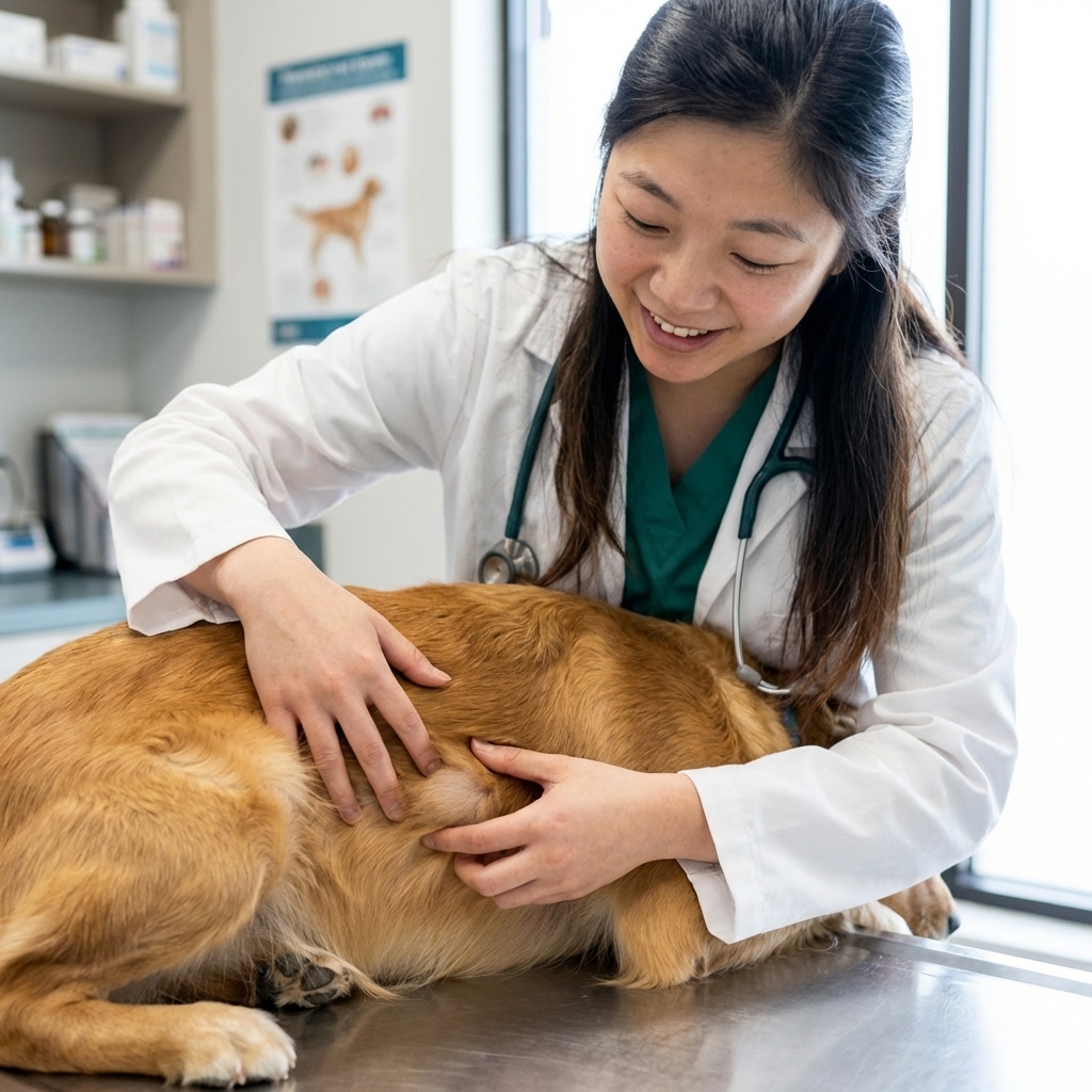
If you have ever been petting your dog and suddenly felt a new lump, you are not alone. In my work as a veterinary assistant here in Frisco, Texas, I see this worry all the time. The good news is that many lumps are benign, including fatty lipomas and cysts. But infections like an abscess and some tumors can look similar at home.
This guide will help you compare the most common “soft lump” culprits by texture, mobility, growth rate, and location, plus a simple checklist you can use at home. I will also tell you exactly when a vet visit is the safest next step.
Important note: At-home checks cannot diagnose a lump. They are meant to help you describe what you are seeing and feeling. The safest rule is still: if it is new, changing, or bothering your dog, get it checked.
Quick definitions
Lipoma
A lipoma is a benign (non-cancerous) tumor made of fat cells. They are very common in middle-aged to older dogs, and they often show up in dogs that are overweight, although fit dogs can get them too.
Cyst (often called “sebaceous”)
Many of the “sebaceous cysts” dog owners notice are actually follicular or epidermal cysts (sometimes called epidermal inclusion cysts). In simple terms, it is usually a blocked hair follicle or skin structure that forms a small sac under or within the skin. These can stay small and quiet, or they can become inflamed or infected.
Abscess
An abscess is a pocket of infection, usually caused by a bite, puncture, foreign material (like a foxtail), or bacteria entering under the skin. Abscesses can look like a sudden lump and often feel warm or painful.
Lipoma vs cyst vs abscess: what you can feel at home
These are the patterns we commonly see when we examine dogs in clinic.
1) Texture
- Lipoma: Usually soft, doughy, or rubbery. Some feel like a squishy “fat pad” under the skin.
- Cyst: Often firmer than a lipoma. May feel like a small marble or pea. If it is close to the surface, it can feel like a dome in the skin.
- Abscess: Can feel swollen and tight at first, then may become more fluid-like as pus collects. Often painful.
2) Mobility (how easily it moves)
- Lipoma: Commonly very movable under the skin. You can often gently wiggle it side-to-side.
- Cyst: May be somewhat movable, but can feel more “anchored” to the skin because it starts in the skin structures.
- Abscess: Mobility varies. The area may feel diffusely swollen rather than a neat, separate ball.
3) Growth rate
- Lipoma: Typically slow-growing over months. Some do grow faster, but a sudden overnight lump is less typical for a lipoma.
- Cyst: Can stay the same size for a long time, then suddenly enlarge if it ruptures or becomes inflamed.
- Abscess: Often appears quickly and can develop over 1 to 3 days, especially after a bite wound or puncture.
4) Location patterns
- Lipoma: Common on the chest, belly, armpits, flanks, and upper legs. They often sit in the fatty layer under the skin.
- Cyst: Often on the trunk, neck, or areas with lots of follicles. Can occur anywhere, including the head.
- Abscess: Common near bite sites (neck, face, legs), or between toes if a foreign body is involved. Location often matches an “entry point.”
5) Skin changes you can see
- Lipoma: Skin usually looks normal over it. Hair coat often unchanged.
- Cyst: May have a visible pore, a small scab, or a thin area of skin. Sometimes a cyst leaks thick white, gray, or yellow material that can look like toothpaste.
- Abscess: Skin may look red or bruised, feel warm, and may start to drain foul-smelling fluid. Your dog may lick it intensely.
Other causes of soft lumps
This is the part I really want you to remember if you are hoping a soft lump “has to be” a lipoma. Dogs can have other soft or squishy masses that look innocent at first glance.
- Mast cell tumors (MCTs): The classic “great imitator.” They can feel soft like a lipoma, and they can also change size from day to day.
- Soft tissue sarcomas: Sometimes feel rubbery and may be more fixed to deeper tissue.
- Enlarged lymph nodes: Can feel like firm or rubbery lumps in typical locations (neck, armpits, groin, behind knees).
- Hematoma or seroma: A blood or fluid pocket after trauma, surgery, or a hard play session.
- Hernia: A soft bulge that may be more noticeable when your dog stands, strains, or barks.
That is why we so often recommend sampling lumps, even when they feel “like fat.”
A simple at-home lump checklist
If you find a new lump, take 3 to 5 minutes to gather information. This helps your vet decide how urgently your dog needs to be seen and what tests are most appropriate.
Step 1: Measure and document
- Measure the lump in two directions (example: 2.0 cm by 1.5 cm). A ruler is fine.
- Take a clear photo from the same angle you can repeat later.
- Write down the date you found it.
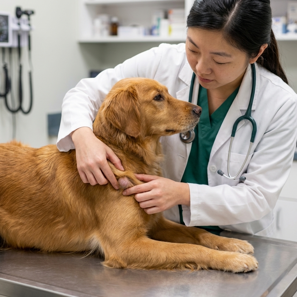
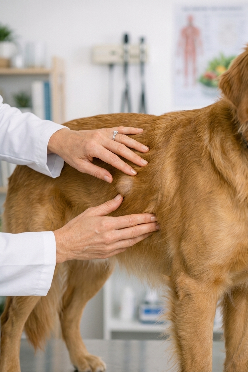
Step 2: Feel (gently) and note what you notice
- Soft and squishy or firm and marble-like?
- Moves freely under the skin or feels stuck?
- Does your dog show pain when you touch it?
- Is the area warm, red, or swollen?
Quick safety tip: Do not keep pressing on it multiple times a day. Excessive palpation can irritate inflamed tissue, and it can really hurt if the lump is infected.
Step 3: Check the surface
- Any pore, scab, or drainage?
- Any odor or wet fur from licking?
Step 4: Watch for whole-body clues
- Normal appetite and energy, or lethargy?
- Any fever signs (warm ears, shivering, acting “off”)?
- Any limping or guarding?
Helpful rule: If a lump is new, growing, changing, or bothering your dog, it deserves a vet visit.
When you should see a vet urgently
Some lumps can wait for a normal appointment, but these should be treated as urgent.
Call your vet within 24 hours (or go in sooner) if:
- The lump appeared very quickly (over hours to a couple of days).
- It is painful, hot, red, or rapidly swelling.
- There is drainage, especially foul-smelling, bloody, or thick fluid.
- Your dog seems unwell: low appetite, lethargy, fever signs.
- The lump is near the eye, mouth, anus, or genitals, where swelling can cause complications.
- The lump is on the foot or between toes and your dog is licking or limping. Foreign bodies and infections love this location.
Book an appointment soon (even if your dog seems fine) if:
- The lump is larger than about 2 cm (about the width of a grape or more) or it is growing. This is a rule of thumb, not a hard cutoff.
- It feels firm, irregular, or fixed to deeper tissue.
- There are multiple new lumps appearing.
- Your dog is a senior, or has a history of tumors.
If you are unsure, call your vet and describe what you found. That is exactly what we are here for.
What your vet will do to tell the difference
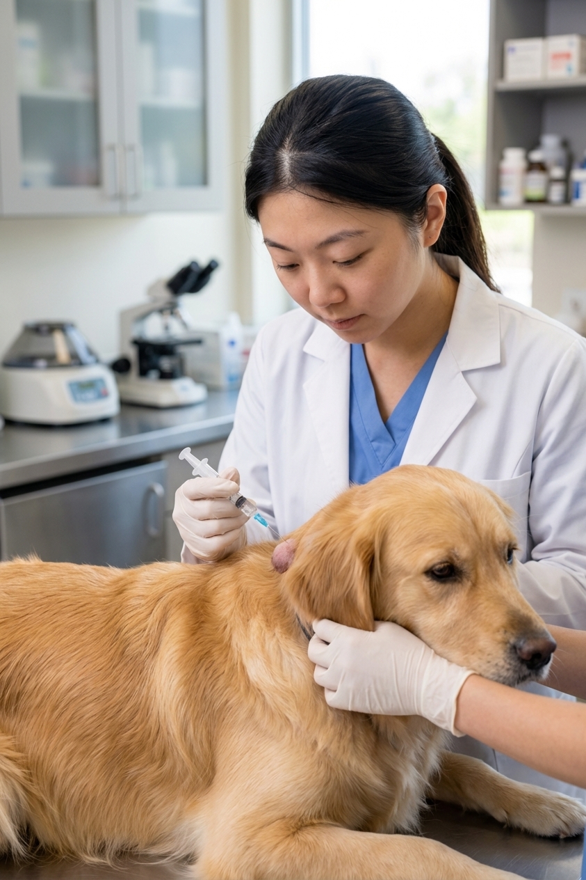
Often, the fastest and most cost-effective first step is a fine needle aspirate (FNA). Your vet uses a small needle to collect cells from the lump, then examines them under a microscope or sends them to a lab.
- Lipoma: FNA often shows many fat cells, which strongly suggests a benign fatty mass.
- Cyst: FNA may pull out oily material, keratin debris, or inflammatory cells, depending on the type and whether it is infected.
- Abscess: FNA often yields pus and bacteria. Your vet may recommend draining, flushing, antibiotics, and pain control, plus checking for a puncture wound or foreign body.
Two quick reality checks that are worth knowing: FNA results can occasionally be non-diagnostic, and the “right” first test can vary by lump type and location. Sometimes a vet may recommend an impression smear (especially if a mass is ulcerated), ultrasound (for deeper masses), biopsy, or surgical removal.
Definitive diagnosis is made with cytology and or histopathology. In other words, even benign-looking lumps can still deserve sampling.
Common myths that can delay care
“If it is soft, it is harmless.”
Many benign lumps are soft, but some concerning growths can also feel soft. Mast cell tumors, in particular, can feel just like a lipoma. Texture alone is not a diagnosis.
“If my dog is not acting sick, it can wait months.”
Some lumps are easiest to treat when they are small. Early evaluation often gives you more options and peace of mind.
“I should squeeze it to see what comes out.”
Please do not. Squeezing can rupture a cyst under the skin, worsen inflammation, or spread infection if it is an abscess. It can also be painful.
“I can lance or drain it at home.”
Please do not do this at home. It can drive infection deeper, cause more swelling, and delay proper treatment.
Safe home care while you wait for your appointment
- Prevent licking: Use an e-collar or recovery collar if your dog is focused on it.
- Keep it clean and dry: If there is mild surface drainage, gently wipe with saline on gauze. Avoid harsh antiseptics unless your vet directs you.
- Do not apply human acne products or essential oils: Many are irritating or toxic if licked.
- Monitor daily: Size, redness, warmth, and your dog’s comfort level.
If anything changes quickly, especially swelling, pain, or your dog seems unwell, bump that appointment up.
The bottom line
At home, lipomas often feel soft and very movable, cysts often feel more firm and skin-attached with possible pore or leakage, and abscesses often show up suddenly with pain, heat, redness, or drainage. But there is overlap, and some tumors (especially mast cell tumors) can mimic benign lumps.
You do not have to panic, but you also do not have to guess. Gather a few notes, take a photo, and let your veterinary team help you get a confident answer.
References
- American College of Veterinary Surgeons (ACVS). Client education resources on skin masses and tumors in dogs.
- Merck Veterinary Manual. Information on skin disorders, abscesses, and benign skin growths in dogs.
- VCA Animal Hospitals. Client education materials on lipomas and follicular epidermal cysts in dogs.