
Horner’s Syndrome in Dogs: Benign or Emergency?

If your dog suddenly looks like they are squinting on one side, with a smaller pupil and a slightly sunken eye, it can be alarming. I get it. As a veterinary assistant, I have seen how fast pet parents go from “Is this just allergies?” to “Is my dog having a stroke?”
Horner’s syndrome is one of those conditions that looks dramatic but the eye changes themselves are rarely life-threatening. That said, Horner’s can be a clue that something is happening deeper in the ear, neck, chest, or even the brain. The key is knowing what to look for and how quickly to get help.
Quick note: This article cannot diagnose your pet. If you are unsure, call your veterinarian or an emergency clinic for guidance.
What it looks like
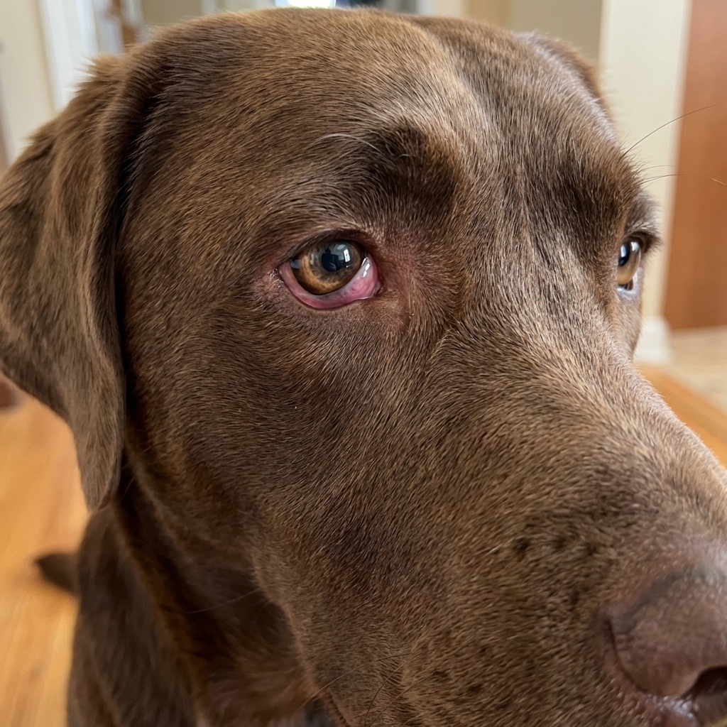
Horner’s syndrome is a set of eye and eyelid changes caused by disruption of sympathetic nerves that normally help control the eye and eyelids.
The classic triad (plus one)
- Small pupil (miosis) on the affected side
- Droopy upper eyelid (ptosis)
- Sunken appearance of the eye (enophthalmos)
- Raised third eyelid (you may see a pale pink membrane partially covering the eye)
These changes usually affect one eye. The eye itself often is not painful with Horner’s, but the underlying cause can be.
Common questions I hear
- “Is my dog going blind?” Usually no. Horner’s changes the look of the eye more than the ability to see.
- “Is it a seizure or stroke?” Horner’s alone is not a seizure. A stroke is possible but less common, and other neurologic symptoms are typically present.
What can look similar
This is important, because a “weird-looking eye” can be Horner’s, but it can also be something that needs urgent treatment.
- Corneal ulcer or eye injury: usually painful, with strong squinting, tearing, and sensitivity to light.
- Uveitis (inflammation inside the eye): can cause a small pupil and squinting, often painful, sometimes with redness.
- Glaucoma: often a very painful eye with a cloudy or blue-tinted cornea, and the pupil is commonly very dilated (but owners can notice “pupil changes” either way).
- Facial nerve paralysis: can cause droopy facial features, but it looks different than classic Horner’s and often includes an inability to blink normally.
If your dog is acting painful, the eye is cloudy, or vision seems suddenly off, treat that as urgent even if you suspect Horner’s.
Why it happens
The sympathetic nerve supply to the eye is like a long relay race. It starts in the brain, travels down the spinal cord, exits in the chest area, runs back up the neck, then runs close to the middle ear (the tympanic bulla) and surrounding tissues before reaching the eye.
Because that route is so long, Horner’s can be triggered by problems in several places. Veterinarians often describe the location as:
- Central: brain or upper spinal cord
- Preganglionic: chest to neck region
- Postganglionic: middle ear area to the eye
This “where is the problem?” question is why your vet may recommend a specific eye test, ear exam, or imaging.
Common causes in dogs
Many cases are linked to the ear or to trauma. Others are harder to pin down. Here are some of the more common possibilities.
Middle ear disease
This is a big one. The nerves involved run close to the middle ear, so infection or inflammation can interfere with them.
Clues can include:
- Recurring ear infections
- Head shaking or ear scratching
- Ear odor or discharge
- Pain when the ear is touched
Inner ear disease and vestibular episodes
Vestibular disease can cause head tilt, loss of balance, and rapid, involuntary eye movements (nystagmus). Horner’s can occur alongside vestibular changes because the middle and inner ear live in the same neighborhood.
If your dog has Horner’s changes plus:
- Head tilt
- Walking like they are drunk
- Vomiting or severe nausea
- Nystagmus
...ear disease is often a leading concern, especially when vestibular changes are part of the picture. Your vet will also keep neurologic causes on the list.
Idiopathic (no cause found)
In some dogs, no cause is found even after a solid workup. These cases often improve over time, but they still deserve an initial exam to be sure you are not missing ear disease or trauma.
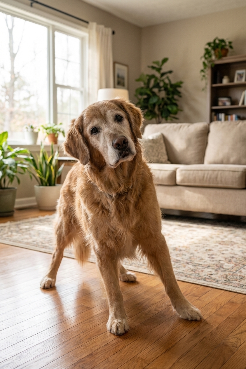
Breed note: Golden Retrievers are known to be overrepresented in idiopathic Horner’s syndrome, meaning they can develop it without a clear underlying cause more often than many other breeds.
Trauma and procedure effects
Because the nerve pathway runs up the neck and near the ear, injuries and some procedures there can trigger Horner’s syndrome.
- Bite wounds around the neck or near the ear can cause swelling, infection, or direct nerve injury.
- Blunt trauma (rough play, falls, car accidents) can stretch or bruise the nerve pathway.
- After ear flushing or deep ear cleaning (sometimes done under sedation or anesthesia), some dogs develop temporary Horner’s changes.
- After ear surgery, Horner’s changes can also occur.
If your dog has any puncture wounds, swelling, pain, or fever, treat this as urgent. Bite wounds can look small on the outside but be significant underneath.
Less common but important causes
Most dogs do not have a tumor or brain lesion, but we do not want to miss the cases that do.
Masses affecting the nerve pathway
Tumors or growths can occur in the:
- Ear canal and or middle ear (tympanic bulla)
- Neck
- Chest (near where nerves travel upward)
Neurologic disease
Problems in the brain or upper spinal cord can sometimes cause Horner’s syndrome, especially if there are other neurologic symptoms (weakness, behavior change, seizures, trouble swallowing).
Benign or emergency?
Horner’s syndrome is often not an “ER right now” diagnosis by itself. But it can be the visible tip of a bigger problem. Here is how I think about it for families.
Go to an emergency vet today if you notice
- Eye pain: pawing at the eye, strong squinting, obvious discomfort
- Red, cloudy, or blue-looking cornea
- Sudden blindness or bumping into things
- A suddenly very dilated pupil (especially with pain or cloudiness), which can be a glaucoma clue
- Head trauma or a known bite wound
- Severe vestibular changes: nonstop falling, vomiting, cannot stand
- Other neurologic symptoms: seizures, severe weakness, collapse, abnormal mentation
Book a prompt vet visit (within 24 to 48 hours) if
- The eye looks “different” but your dog is otherwise comfortable
- No trauma is known
- Your dog has ear infection history, head shaking, or ear odor
Even when it is not an emergency, an exam matters because untreated ear disease can worsen and become very painful.
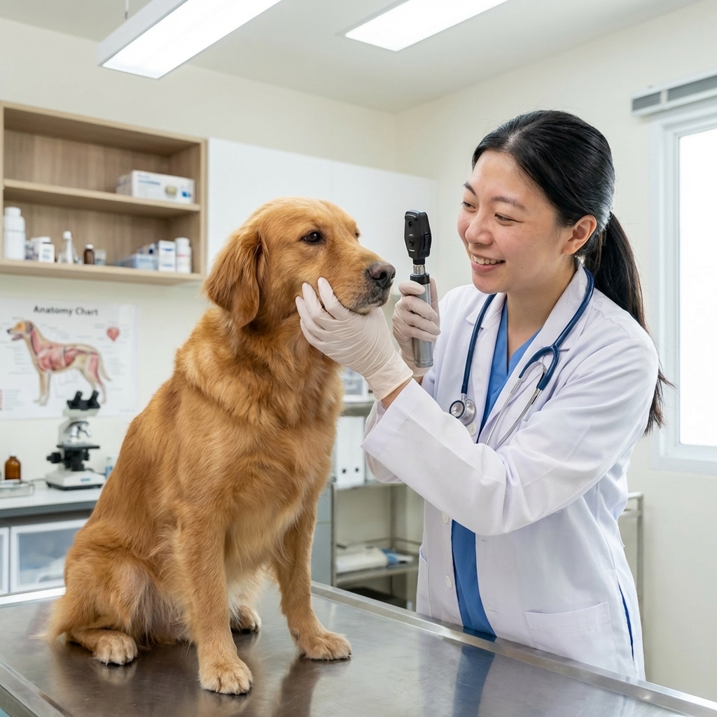
What your vet will do
Most appointments start with two goals: confirm it is Horner’s syndrome and look for the underlying cause.
1) Physical and neurologic exam
Your vet will check:
- Eye appearance and pupil response
- Eyelid position and third eyelid
- Any facial asymmetry or pain
- Gait, balance, and reflexes
2) Eye tests to protect the cornea
- Fluorescein stain to look for corneal ulcers
- Tear testing if dry eye is suspected
- Intraocular pressure to help rule out glaucoma (glaucoma is painful and urgent)
These are quick tests that help rule out problems that can threaten vision.
3) Ear exam
Your vet may do an otoscopic exam and may recommend:
- Ear cytology (looking at debris under a microscope)
- Ear culture for complicated or recurrent infections
If the ear canal is too inflamed or painful to examine deeply, sedation may be recommended for a safe, thorough look.
4) A classic eye drop test (sometimes)
In some cases, your veterinarian may use a phenylephrine eye drop test. It can temporarily reverse Horner’s changes, and the timing of that improvement can help localize where along the nerve pathway the problem may be. Not every clinic uses this test, and it is not required to treat many cases, but it is a fascinating tool when it fits.
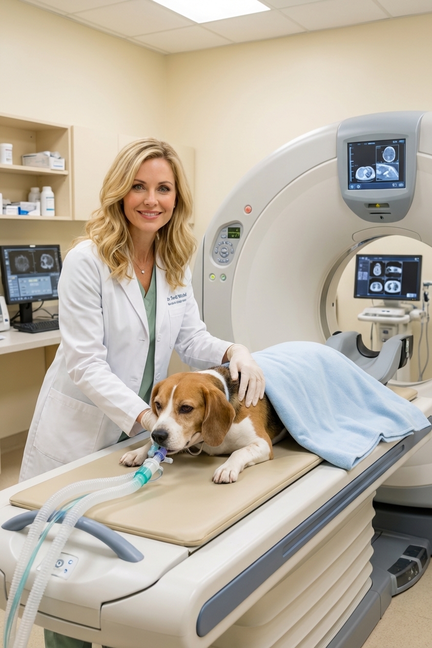
5) Imaging
Imaging is not always needed, but it becomes more likely when:
- There are vestibular changes
- Middle or inner ear disease is suspected
- Changes persist or worsen
- Your dog has other neurologic abnormalities
- A mass is suspected
CT is often excellent for evaluating bony structures like the ear canal and middle ear (tympanic bulla). MRI is best for soft tissue and brain. Your vet will choose based on the most likely cause and what is available.
How it is treated
There is no single “Horner’s medicine.” Treatment focuses on the cause, plus keeping your dog comfortable.
If ear disease is the cause
- Prescription ear medication (antibiotic, antifungal, anti-inflammatory depending on cytology)
- Pain relief if needed
- Sometimes oral medications for deeper infections
- Recheck visits to confirm the infection is truly clearing
If trauma is the cause
- Pain management
- Wound care and antibiotics if indicated
- Monitoring for improvement
If no cause is found
Many idiopathic cases improve over time. A common timeframe is about 4 to 8 weeks, but some dogs take longer. Your vet may recommend a watch-and-wait plan with rechecks. If changes worsen or new symptoms appear, the plan shifts toward imaging and deeper diagnostics.
What you can do at home
While you are waiting for your appointment or after your visit, these steps are genuinely helpful.
Do
- Take clear photos of your dog’s face daily in the same lighting to track change
- Check for ear symptoms: odor, discharge, head shaking, pain
- Prevent rubbing if your dog is pawing at the eye (ask your vet if an e-collar is appropriate)
- Give medications exactly as prescribed, especially ear meds. Stopping early is a common reason infections come back.
Do not
- Use leftover eye drops from an old problem unless your vet approves
- Put peroxide or alcohol in ears
- Delay care if there is pain, cloudiness, trauma, or balance problems
Prognosis
Many dogs do very well, especially if the underlying issue is treatable and addressed early.
- Idiopathic cases often improve over time.
- Ear infection related cases can improve once the infection and inflammation are controlled, though it may take weeks.
- Trauma related cases vary. Some resolve fully, others improve partially.
- Masses or neurologic causes depend on what is found and how early it is treated.
One encouraging note: even when the eye looks odd, many dogs feel normal and behave normally. Your job is to make sure we are not missing the condition behind the change.
Quick recap
Horner’s syndrome is a cluster of changes like a small pupil, droopy eyelid, and sunken eye, usually on one side. It is often linked to ear disease or trauma, can overlap with vestibular episodes, and should be checked by a veterinarian promptly. Seek emergency care if there is eye pain, cloudiness, trauma, severe balance issues, a suddenly very dilated pupil, or other neurologic symptoms.