
Histiocytoma in Dogs: The Common Red Button Bump

If you have ever felt a small, sudden “button” on your dog’s skin and your stomach instantly dropped, you are not alone. One very common cause of a fast-appearing, red, hairless bump in younger dogs is a histiocytoma. The reassuring news is that histiocytomas are benign (non-cancerous) skin tumors, and many resolve on their own.
That said, some dangerous skin tumors can look similar at home, especially mast cell tumors. So the goal of this guide is to help you recognize the typical pattern of a histiocytoma, understand what else it could be, and know exactly when it is time to see your veterinarian.
What is a histiocytoma?
A histiocytoma is a benign skin tumor that forms from immune system cells in the skin called histiocytes (often described as dendritic cells). In plain language, it is a small overgrowth of certain “immune” cells in the skin.
They are especially common in:
- Young dogs, typically under 3 years
- Dogs with a strong immune response (many histiocytomas regress because the immune system recognizes and clears them)
- Any breed, though they are often reported in breeds like Boxers, Boston Terriers, Dachshunds, and some retrievers and terriers
Even though histiocytomas are benign, you should still treat any new lump as “needs identification,” because look-alikes exist.
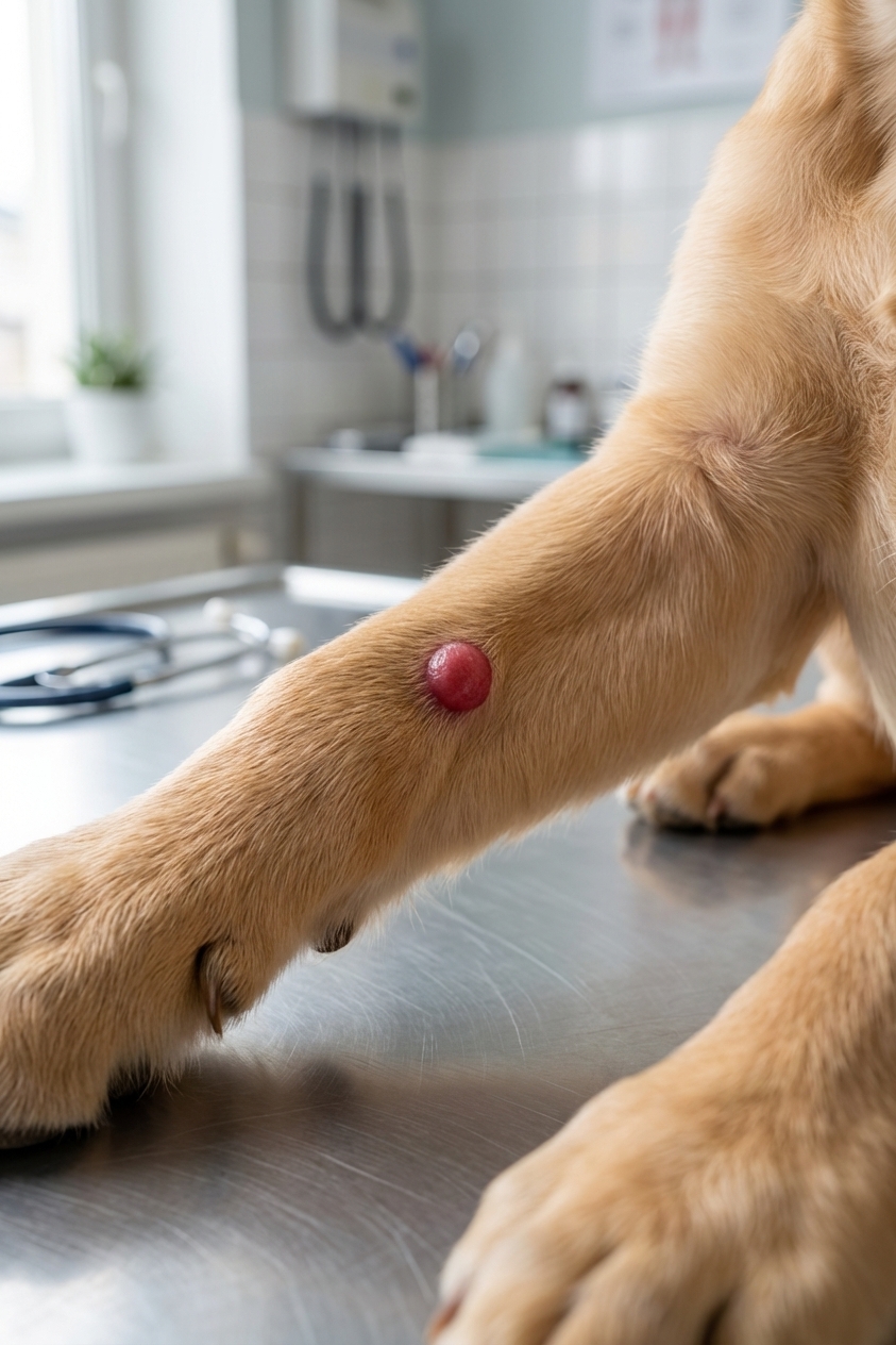
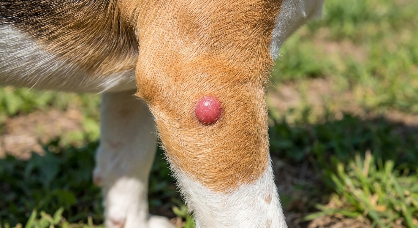
What a histiocytoma looks like (the classic “red button”)
Owners often describe histiocytomas as if a little pink or red button popped up overnight. Typical traits include:
- Round and well-defined
- Raised, like a dome
- Hairless on top (or very thin-haired)
- Pink to bright red
- Usually small (often pea-sized, but can be larger)
- Commonly found on the head, ears, neck, and limbs
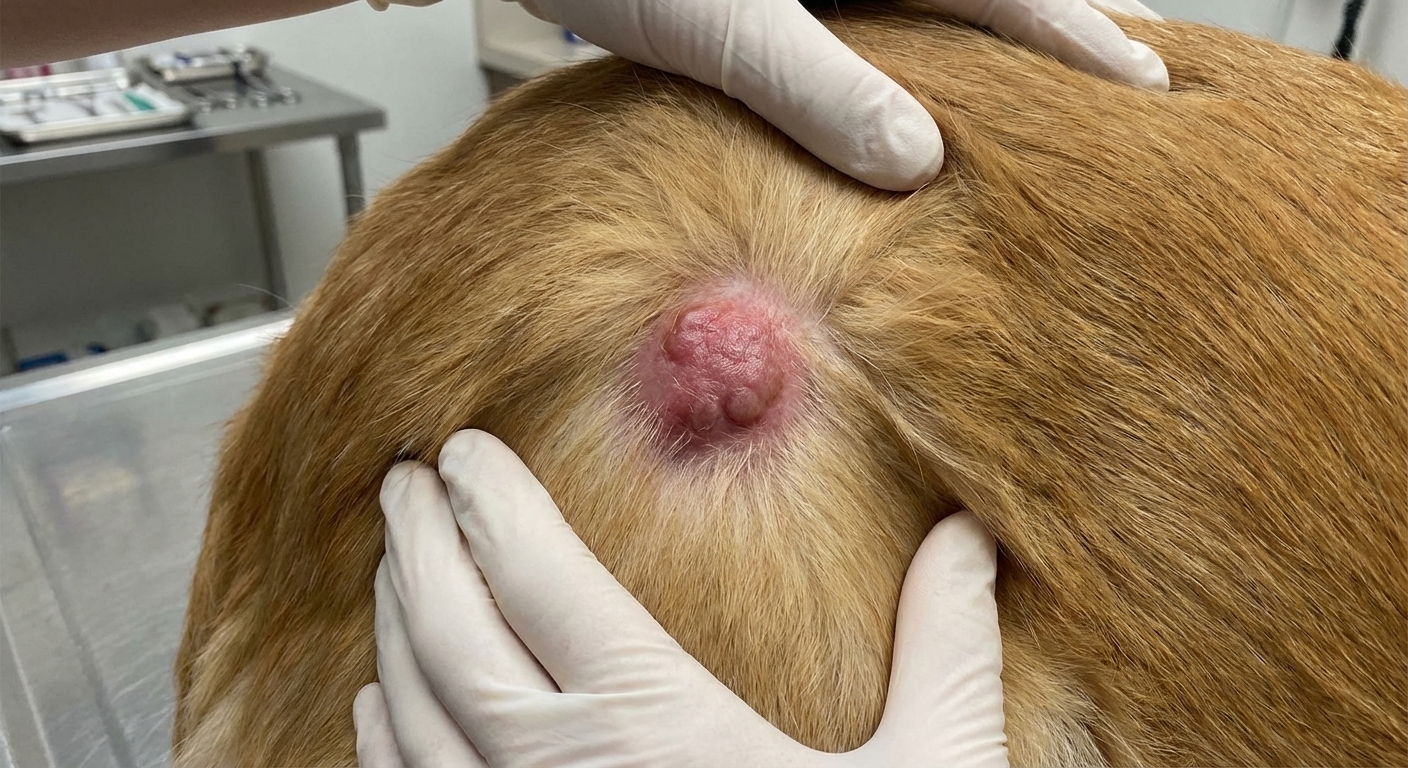
Many are not painful, but they can become itchy or irritated. Some ulcerate (get a raw spot) because the dog licks or scratches them.
Histiocytoma vs mast cell tumor vs wart
Here is the tricky part: at home, you cannot reliably diagnose a skin tumor by appearance alone. Still, the pattern can help you understand what your vet may be considering.
Histiocytoma vs mast cell tumor (the important one)
Mast cell tumors can mimic almost anything: they might be pink, red, skin-colored, raised, flat, ulcerated, small, or large. Some grow quickly. Some change size from day to day due to histamine release and inflammation. Because of this, vets often recommend a fine needle aspirate (FNA) to check cells under a microscope.
- Histiocytoma tendency: younger dog, classic red button look, often regresses over weeks
- Mast cell tumor tendency: any age (more common in middle-aged to older dogs), may swell, shrink, or look angry and inflamed, can be itchy, can be solitary or multiple
If your dog is not a young pup or young adult, or the bump looks “weird,” it is extra important to get it checked promptly.
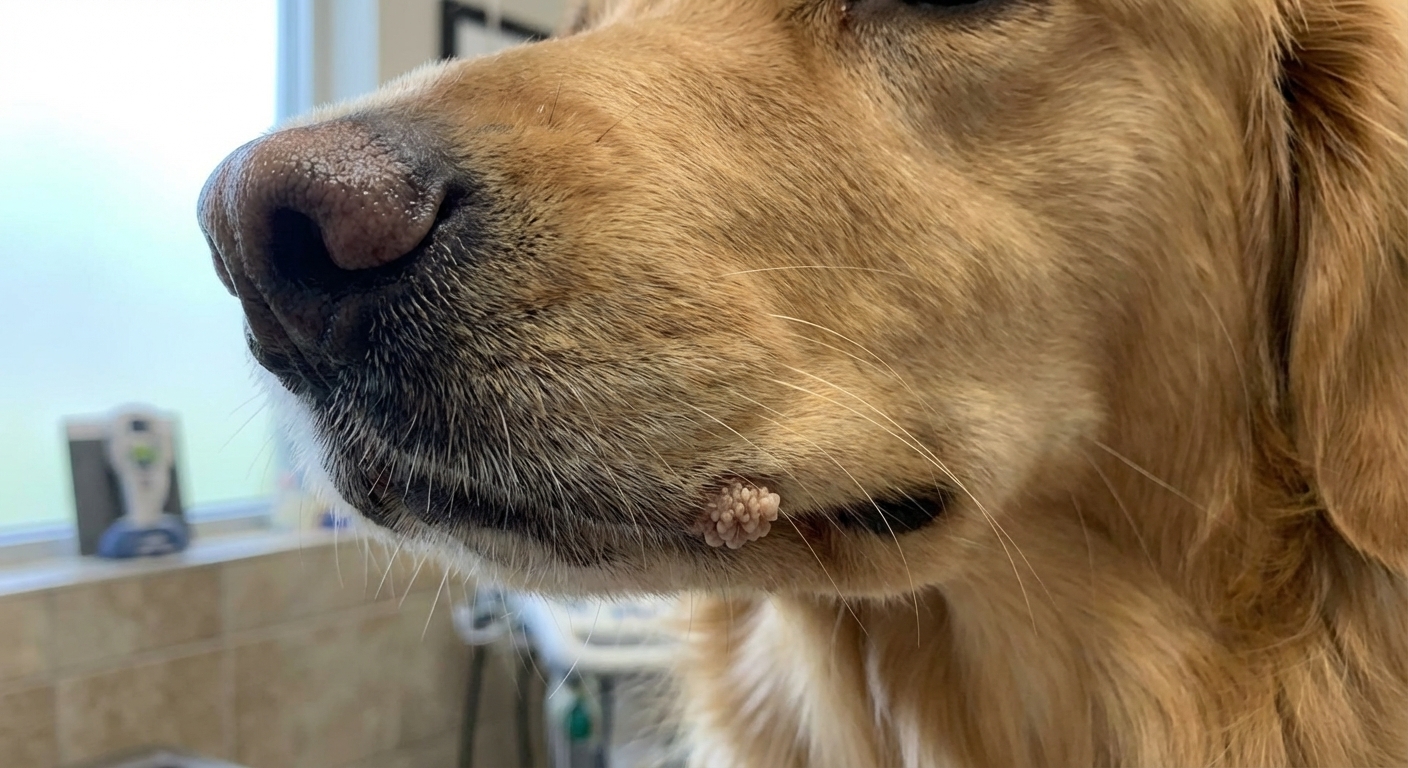
Histiocytoma vs papilloma (wart)
Canine papillomas are warts caused by a virus and are common in young dogs too. Warts often look:
- Cauliflower-like or frilly on the surface
- Grouped or multiple (often around the mouth, lips, eyelids, or paws)
- More textured than the smooth “button” style
Histiocytomas are usually smoother and more dome-shaped.
Typical progression and how long they take to go away
A very common histiocytoma timeline looks like this:
- Week 1 to 2: a small red bump appears and may grow quickly
- Weeks 2 to 6: the bump may stay the same size, look more irritated, or develop a small scab if your dog licks it
- Weeks 4 to 12: many begin to shrink and flatten as the immune system clears it
Many histiocytomas resolve within 2 to 3 months. Some resolve sooner, and some take longer. If it is truly a histiocytoma, “watchful waiting” is often reasonable, but only after your veterinarian has confirmed the diagnosis.
Important: do not assume a lump is a histiocytoma just because your dog is young. Your vet can often confirm with an FNA, which is quick and typically does not require sedation.
When surgical removal is recommended
Because histiocytomas often self-resolve, many do not require surgery. Still, removal can be the best choice in certain situations.
Your vet may recommend removal when:
- The diagnosis is uncertain (FNA is inconclusive, or the appearance is atypical)
- The mass is rapidly enlarging or changing in an unusual way
- It ulcerates, bleeds, or becomes repeatedly infected
- Your dog cannot stop licking, chewing, or scratching it
- It is in a spot that causes problems, like the eyelid, lip, ear margin, paw, or between toes
- It has not improved after a reasonable watch period (often around 8 to 12 weeks, guided by your veterinarian)
Surgical removal is typically straightforward, and the tissue is often sent to a lab for histopathology to confirm what it is. That confirmation matters, because mast cell tumors and other growths can require wider surgical margins and different follow-up.
What you can do at home (safe, helpful steps)
Here are practical steps I recommend to families when a new bump appears:
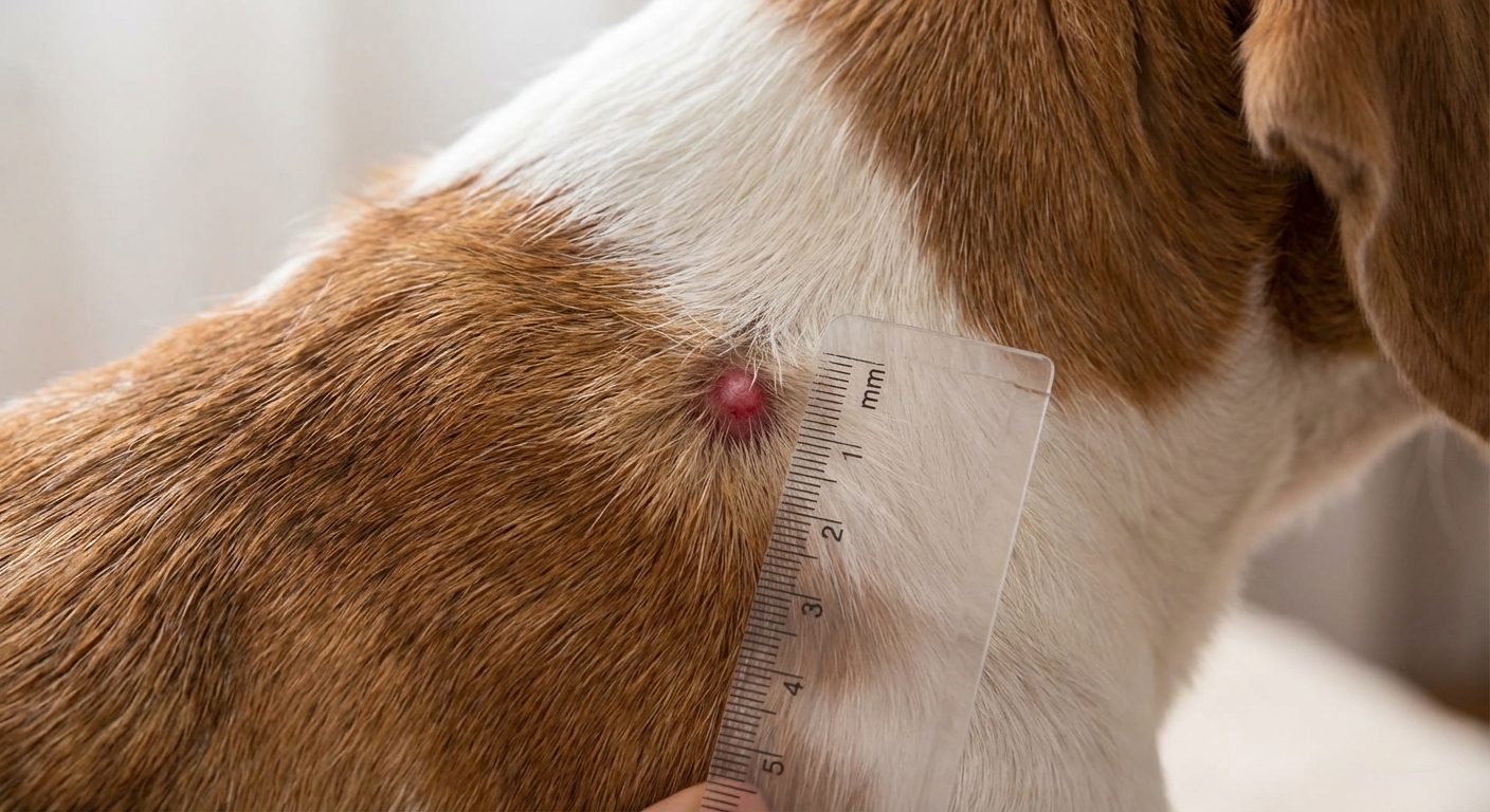
- Measure it with a ruler and write it down (millimeters matter).
- Take a clear photo once a week in the same lighting and distance.
- Prevent licking if it is getting raw. An e-collar can protect the area while you wait for your appointment.
- Do not apply human creams unless your veterinarian tells you to. Some products can irritate skin or be unsafe if licked.
- Book a vet visit for any new lump that lasts more than a week, grows quickly, bleeds, or looks infected.
Trust your instincts. If a bump is changing fast, looks angry, or just does not seem “right,” it deserves a closer look.
When to go to the vet urgently
Most histiocytomas are not emergencies, but you should contact your veterinarian promptly if you notice:
- Sudden swelling around the lump, hives, vomiting, diarrhea, or weakness
- Rapid growth over days
- Bleeding that will not stop, deep ulceration, or pus-like discharge
- A lump on the face that interferes with an eye, or a lump between toes causing limping
These signs do not guarantee cancer, but they do mean “do not wait and see.”
Photo guide: what to capture for your vet
If you can snap a few good photos, it can genuinely help your clinic track changes.
- Full-body context photo: where the lump is located on your dog.
- Close-up photo: fill the frame with the lump, in focus.
- Size reference: a ruler placed next to the lump (do not press on it).
The bottom line
Histiocytomas are one of the most common benign skin tumors in young dogs. They often look like a round, raised, hairless, red button and many resolve on their own within weeks to a few months. The catch is that other skin tumors, including mast cell tumors, can look similar, so a quick vet check and an FNA can bring peace of mind and prevent delays if the lump is something more serious.
If you are unsure, take photos, measure the bump, and schedule a visit. You are not overreacting. You are being a great advocate for your dog.